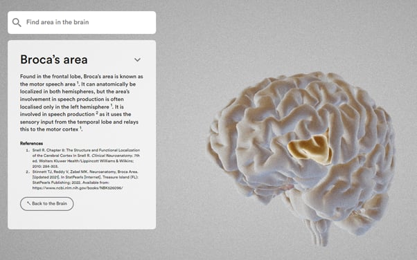
Review the brain from a 360-degree viewpoint
The Brain Atlas allows you to examine structures of the brain in accurate 3D imaging from a 360-degree viewpoint. Use the mouse to navigate all around from top to bottom, from side to side, and to zoom in on relevant structures. By clicking on a desired structure or region, the drop-down bar on the left side will present the name of the structure as well as a detailed description of its anatomical and functional qualities.
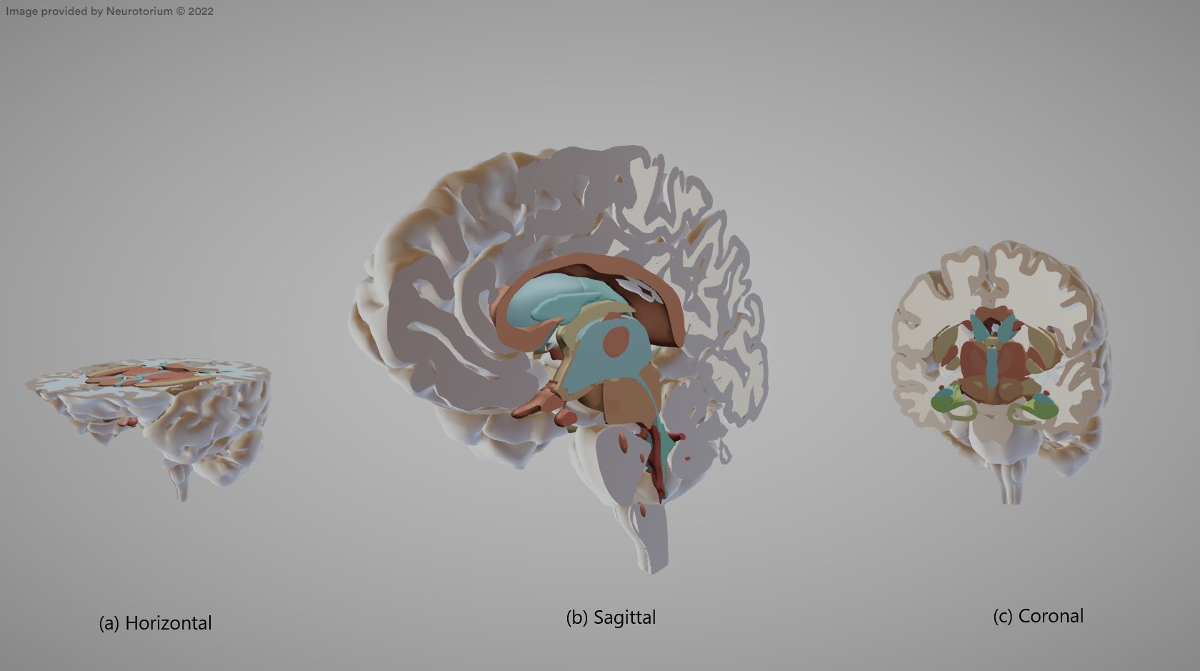
Move slice plane
The Brain Atlas provides a feature allowing users to generate sagittal, horizontal, and coronal incisions through the slice plane tool in the right-hand side bar (Illustration 2). Illustration 2b presents the brain from a sagittal incision commonly used in modern imaging techniques.
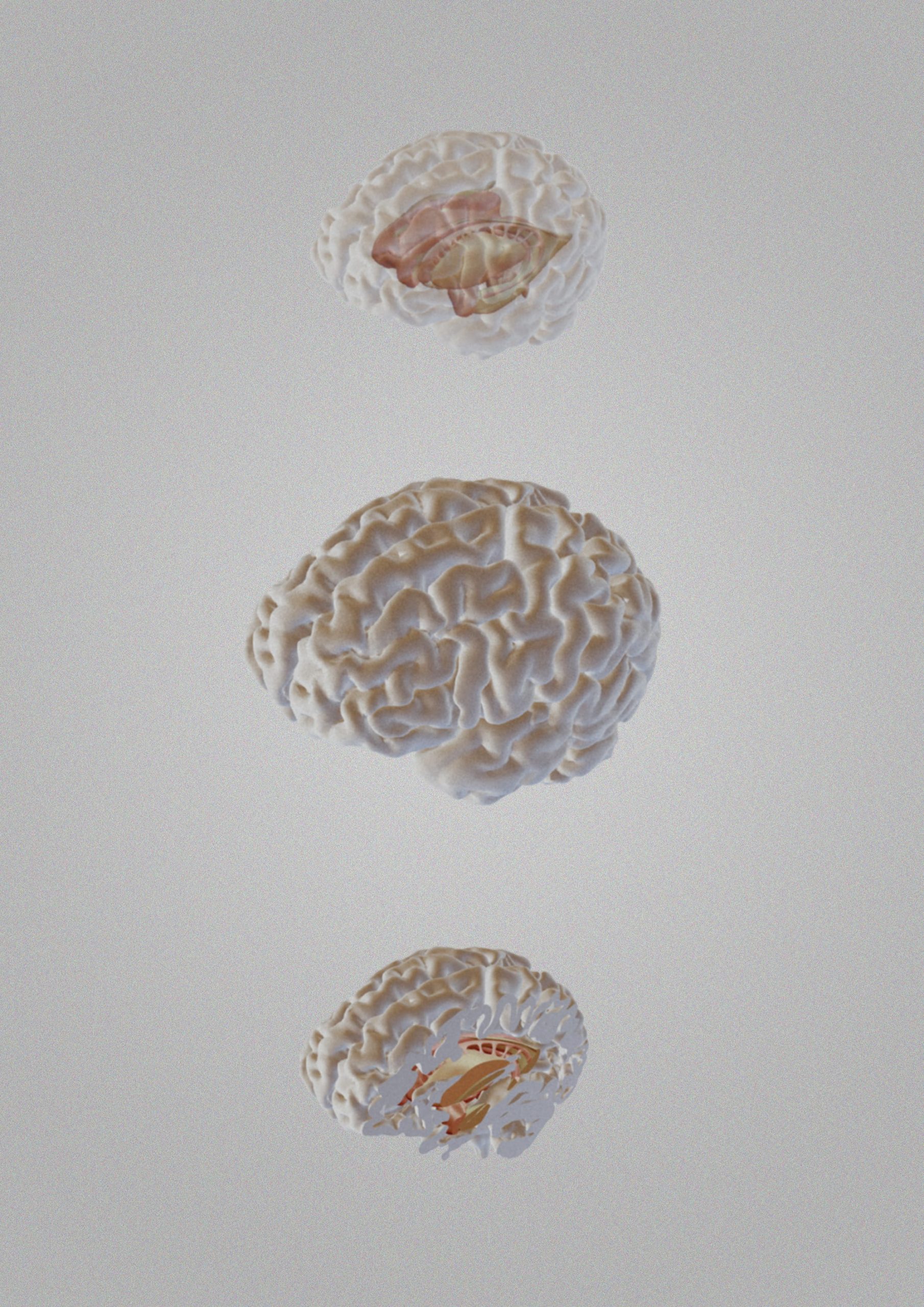
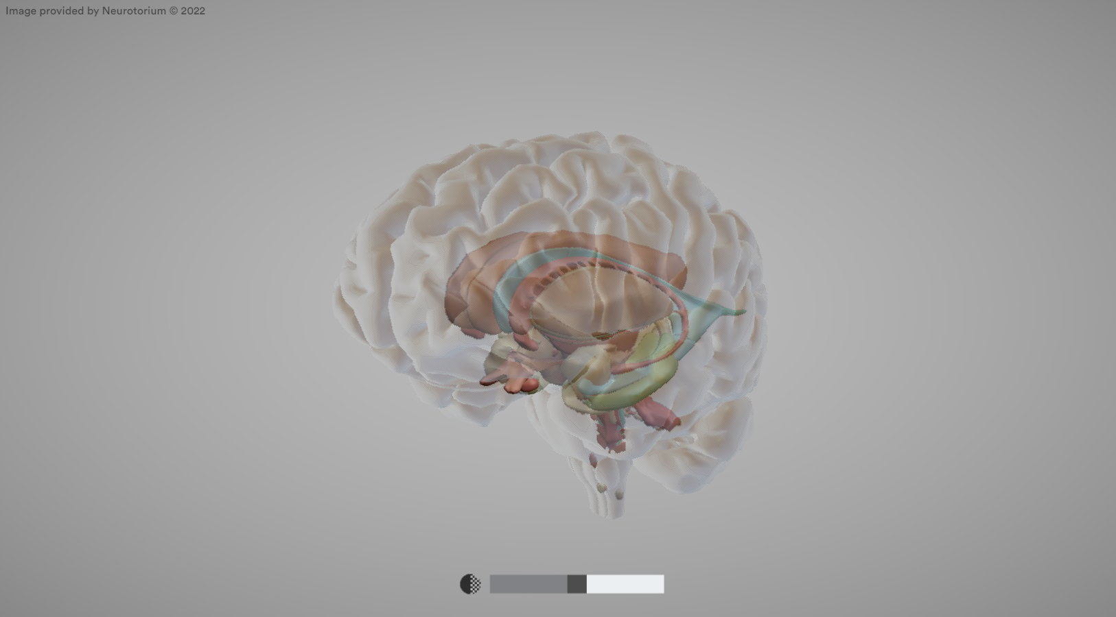
Set surface opacity
Additionally, users can adjust the surface opacity, resulting in the cortex being rendered more or less transparent, and thus revealing subcortical structures, such as the basal ganglia, hippocampus, amygdala, and ventricles. Use the up arrow key to increase the surface opacity or the down arrow key to decrease it.
These tools hold great promise for advancing our academic understanding of the anatomical features of the brain.
Download useful images
Once you have found a desired structure or region of the brain shown at a useful angle and level of transparency, the image can be downloaded for personal use, for teaching, or for your own presentations. Thus, the Brain Atlas contributes to new 3D educational insight into the structures of the brain.
The 3D brain atlas has been developed for Neurotorium, by Random42 and Martin Fredensborg Rath, Professor of Experimental Neuroanatomy, University of Copenhagen and reviewed by the Neurotorium Editorial Board.
Related content

Summary of the physiological changes associated with reproductive aging and the potential links between the menopausal transition and mental health conditions.
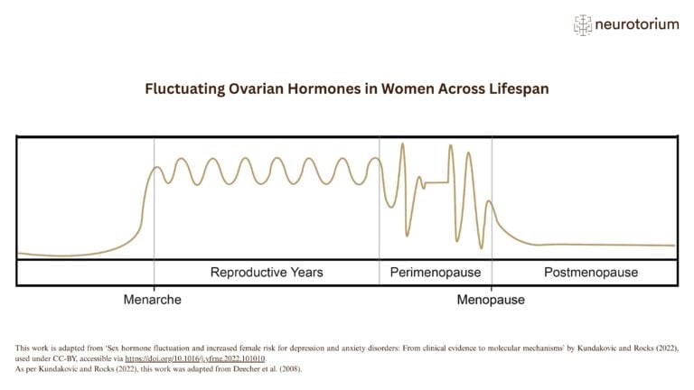
This illustration shows key phases of ovarian hormone fluctuation across the female lifespan, from menarche to menopause.
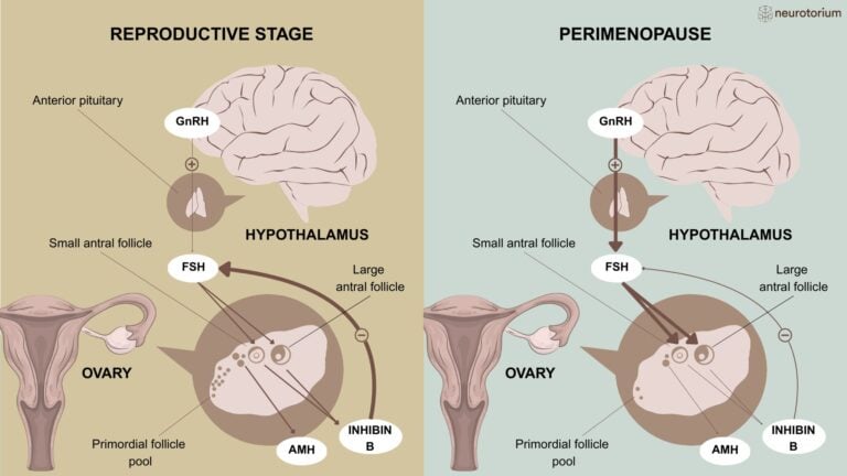
Physiological changes in endocrine function from the reproductive stage to perimenopause