
There are often differences between anterior, middle, and posterior cerebral artery infarctions.1,2
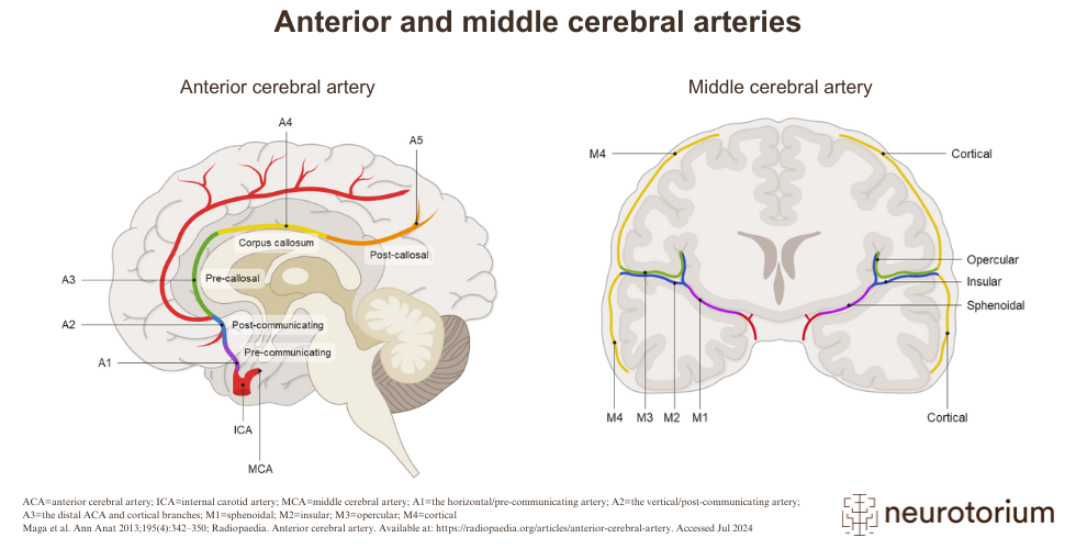
The anterior circulation refers to the part of the brain supplied by the carotid arteries, separated into parts:2,3
- A1: the horizontal/pre-communicating artery
- A2: the vertical/post-communicating artery
- A3: the distal anterior cerebral artery (ACA)and cortical branches
The middle cerebral artery (MCA) is the territory most commonly involved in acute stroke.1,4
The MCA is subdivided into four branches, which supply blood to parts of many lobes of the brain as well as deeper structures such as the caudate and thalamus:4,5
- M1: sphenoidal
- M2: insular
- M3: opercular
- M4: cortical
Related content

Lacunar infarctions are small subcortical infarcts, <1.5 cm in diameter, within deep areas of the brain

The cerebellum is supplied with blood via three branches of the basilar artery – the posterior inferior cerebellar artery (PICA), the anterior inferior cerebellar artery (AICA), and the superior cerebellar artery (SCA).

The image shows how the blood supply of the brain is arranged.