
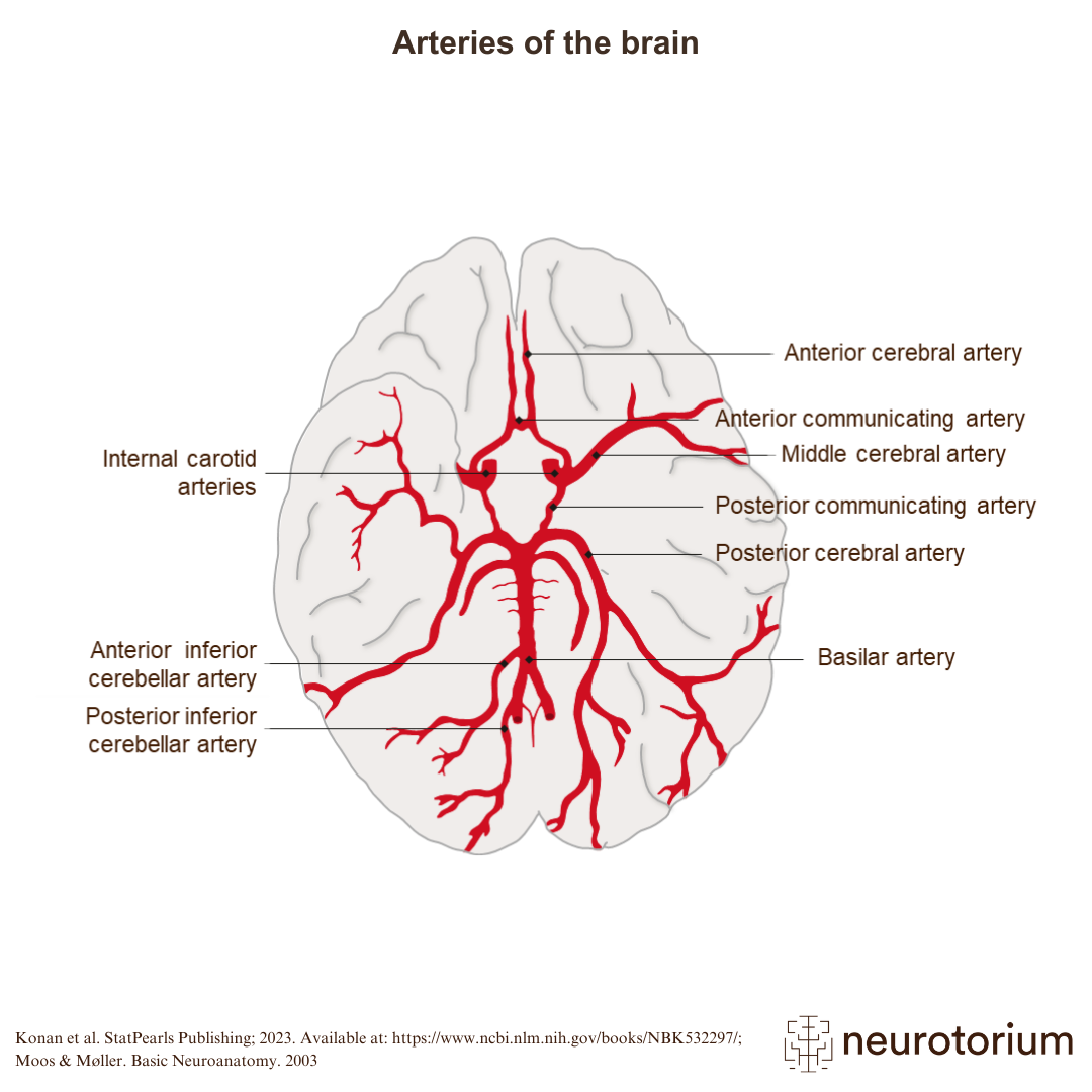
The image shows how the blood supply of the brain is arranged.1,2 The internal carotid arteries branch to form two major cerebral arteries on each side:1,2
- The anterior cerebral artery (ACA): supplies medial frontal and parietal lobes
- The middle cerebral artery (MCA): supplies lateral frontal, parietal, and temporal lobes
The posterior circulation to the brain is mediated by the vertebral arteries which join at the level of the pons to form the basilar artery, which branches into the right and left.1,2
The posterior cerebral arteries (PCA) supply the occipital lobe and temporal lobe.1-3
Related content

Lacunar infarctions are small subcortical infarcts, <1.5 cm in diameter, within deep areas of the brain

The cerebellum is supplied with blood via three branches of the basilar artery – the posterior inferior cerebellar artery (PICA), the anterior inferior cerebellar artery (AICA), and the superior cerebellar artery (SCA).

There are often differences between anterior, middle, and posterior cerebral artery infarctions.