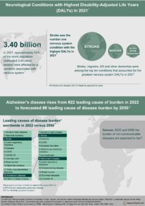
If dementia care were a country, it would be the world’s 18th largest economy.1 The number of people living with dementia today exceeds the entire population of Spain, and this number is projected to almost triple by 2050. Worldwide, dementia costs more than USD 800 billion annually (estimated for 2015). This is more than 1% of global GDP. In the US alone, the USD 109 billion direct health expenditure on Alzheimer’s Disease (AD; i.e., excluding the costs of informal family care) exceeds that on heart disease (USD 102b) and cancer (USD 77b). In short, the global burden of AD, in terms both of human suffering and in costs to society, is immense — and growing.
“In both autosomal dominant and sporadic AD, early increases in cortical thickness in asymptomatic subjects are followed by decreases with emergence of clinical disease”
In unraveling the complex etiology of the disorder, and in the search for biomarkers, it would be helpful to find a clear association – for example — between beta-amyloid in the CSF and loss of cortex in areas relevant to AD. However, a cross-sectional study of 17 healthy elderly controls and 16 people with subjective memory complaints (but preserved cognitive function on testing) suggested a non-linear relationship between CSF beta-amyloid 1-42 and cortical thickness.2 In cognitively preserved subjects with beta-amyloid levels in the middle-tertiles – who were judged to be in a transitional state — cortical thickness in the temporoparietal and precuneus regions, which are areas vulnerable to loss in AD, were thicker than in subjects with normal CSF levels.
Other non-linear trends
The complex evolution of potential neuroanatomical markers of pathology is shown by work on patients with familial AD who carry the presenelin-1 (PSEN1) mutation. Compared to controls, PSEN-1 mutation carriers who were symptomatic showed widespread cortical thinning, especially in the precuneous and pariotemporal regions.3 However, mutation carriers who were asymptomatic had greater cortical thickness in these areas (as well as larger caudate volumes) than controls. As an explanation for this unexpected finding, investigators suggested a possible role for reactive neuronal hypertrophy or inflammatory processes.
Subsequent work by this research group from the Barcelonaβeta Brain Research Center, which confirmed non-linear trajectories of changes in brain structure in familial AD, reinforced their view that a period of neuroinflammation and/or accumulation of amyloid, may precede neurodegeneration.4 At baseline, symptomatic mutation carriers had decreased cortical thickness and volume of cortical and subcortical structures when compared with controls. And, in longitudinal MRI studies, they showed accelerated loss. However, asymptomatic carriers (who were studied a mean of 16 years before expected onset of symptoms) showed greater baseline cortical thickness and volume in temporoparietal regions than controls.
APOE ε4 genotype influences CSF YKL-40 concentrations and brain structure
Apolipoprotein E (ApoE) is centrally involved in neuroinflammation, prompting investigation of the potential role of the APOE ε4 genotype in modulating markers of astroglial activation in preclinical carriers and those with mild cognitive impairment due to AD.5 Higher levels of CSF YKL-40 – which has been linked to the cerebral regulation of astroglia — in both presymptomatic and mildly symptomatic carriers suggested increased astroglial activation even when AD-related biomarkers were not significantly different from controls. Importantly, APOE ε4 carriers showed smaller gray matter volume as YKL-40 levels increased while the reverse was true in non-carriers.
The potential relevance of inflammation is reinforced by work showing that raised levels of another inflammation-related CSF marker linked to AD – sTREM2 – are associated with increases in gray matter volume in patients with early stages of the disease.6 After controlling for factors such as age and p-tau, CSF sTREM2 levels were positively correlated with gray matter volume in the bilateral inferior and middle temporal cortices, precuneus and supramarginal and angular gyri. The authors interpret this as indicative of brain swelling, perhaps as part of an inflammatory response to early neurodegeneration. Whether or not this response is neuroprotective remains to be seen.
Prevention is the wider context
Such research into pre-clinical features associated with AD takes place in the context of a suggested move towards diagnosis based not on symptoms and signs but on biomarkers – notably amyloid and tau – which appear to be “proxies of pathology”. Thus the National Institute on Aging and Alzheimer’s Association Research Framework suggests a definition of AD based on a biological rather than clinical construct.7 Its authors acknowledge that at this stage the proposal is not for use in clinical practice, and that it remains a framework open to discussion and revision. However, there is evidence – for example – that a user-friendly index combining CSF amyloid and tau discriminated between AD patients and healthy controls with acceptable sensitivity and a specificity above 85% when trialled across four European memory clinics.8
“A developing idea of AD is one that allows disease to be present in the absence of clinical symptoms”
The impetus behind research into prodromal and early AD is also an acknowledgement that – in the absence of curative or disease-modifying therapies – efforts at prevention offer an opportunity to combat the growing epidemic of neurodegenerative disease in an aging world.9 Prevention may take the form of encouraging lifestyle changes that appear to reduce risk, or of potential drug treatments that may counter developing AD pathology in the pre-clinical and mildly symptomatic phases of the disease continuum.10
Take home messages
- AD is now defined as a biological construct that reflects the underlying pathology manifesting itself through a clinical continuum ranging from normal cognition to dementia
- Early increases of cortical thickness are followed by decreases, in both autosomal dominant AD and sporadic AD
- AD brain structural changes show a non-linear pattern
- Inflammatory mechanisms linked to astroglial and microglial response affect areas adjacent areas to those affected by neurodegeneration
- High YKL40 levels are associated with longitudinal atrophy while low sTREM levels are associated with both increases and decreases in cortical thickness
Access a collection of articles on Inflammation and Brain Disorders
Acknowledgement
We would like to thank Professor José Luis Molinuevo (Barcelonaβeta Brain Research Center, Barcelona, Spain) for sharing his expertise and insights surrounding inflammation in Alzheimer’s disease and for providing feedback in the development of this article.


