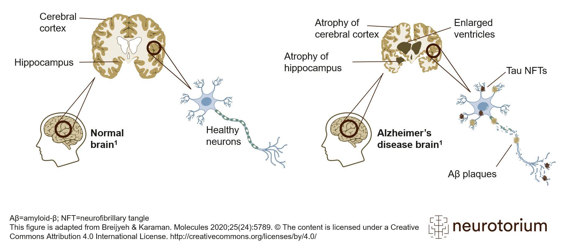
Schematic representation of healthy brain (left) and Alzheimer’s disease (AD) brain (right). Compared to the healthy brain, the brain with AD, as the disease progresses, features an atrophied cerebral cortex, shrinkage of the hippocampus, and enlargement of the ventricles. The AD brain has two other key features: amyloid-beta plaques and tau neurofibrillary tangles (NFTs). Aβ plaques are located extracellularly, and tau NFTs are located intracellularly. These two pathologies are the defining features of AD and are required for a neuropathological diagnosis of AD. This figure is adapted from Breijyeh & Karaman. Molecules 2020;25(24):5789.
The content is licensed under a Creative Commons Attribution 4.0 International License. https://creativecommons.org/licenses/by/4.0/
Related content
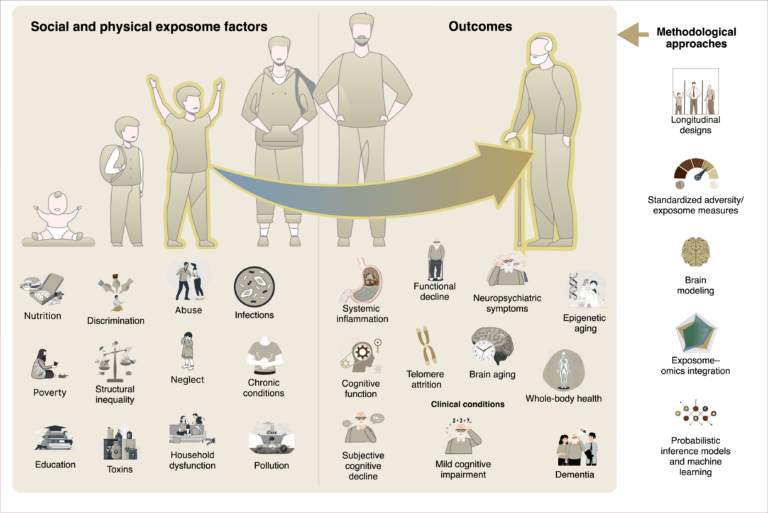
Social and physical exposome factors—like poverty, pollution, and inequality—accumulate over time, shaping biological aging and cognitive decline. Through inflammation and cellular stress, these exposures drive neurodegeneration, highlighting the need for longitudinal, integrative research.

The exposome reframes dementia as the product of cumulative physical and social exposures-like pollution, nutrition, and inequality-across life. These factors shape brain aging through inflammation and epigenetic change. Lifespan, global studies are key to equitable dementia prevention.