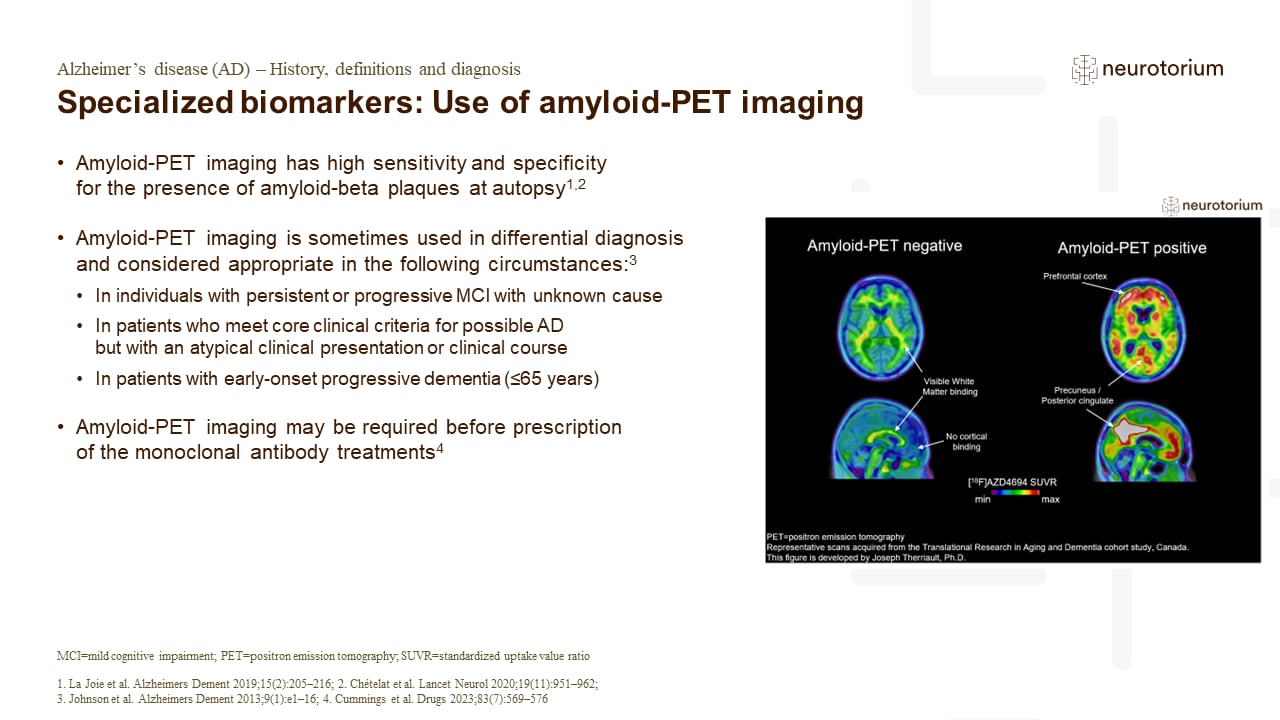
Figure: Amyloid-PET imaging in Alzheimer’s disease: Examples of amyloid-negative (left) and amyloid-positive (right) positron emission tomography (PET) scans.5 In this figure, the amyloid-PET ligand [18F]AZD4694 is shown.5 The hotter colors indicate more amyloid-PET tracer binding, and colder colours indicate low amyloid-PET tracer binding.5 The prototypical amyloid-positive PET pattern is of elevated amyloid in the brain’s posterior cingulate cortex, prefrontal cortex, as well as lateral parietal and temporal cortices.5 These regions display minimal tracer uptake in the amyloid-negative case (left), who has low binding across the whole cerebral cortex.5 Finally, it is important to note that amyloid-PET tracers have some binding to white matter, which explains the small amounts of hotter colours in the amyloid-negative case.5 More information on amyloid-PET visual reads can be found in Johnson KA, Minoshima S, Bohnen NI, et al. Alzheimers Dement. 2013;54(3):476-490.5
Amyloid-PET uses standardized tracer-specific visual reading procedures and has documented high reproducibility across PET centres.2 As a neuroimaging technique, it allows non-invasive, in vivo detection of amyloid plaques, one of the key pathological characteristics of AD.2 The prototypical amyloid-positive PET pattern is of elevated amyloid in the brain’s posterior cingulate cortex, prefrontal cortex, as well as lateral parietal and temporal cortices.5 These regions display minimal tracer uptake with low binding across the whole cerebral cortex in patients that are amyloid negative.5 Amyloid-PET can also allow the identification of amyloid pathology in clinically atypical variants of AD including posterior cortical atrophy, frontal-executive variant, or the logopenic variant,2,6,7 however, it does not provide the ability to distinguish between distinct amyloid-positive disorders which may show similar patterns of amyloid deposition.2 It should be noted that amyloid-PET tracers have some binding to white matter, resulting in small amounts of hotter colours observed on tracers in amyloid-negative cases.5
When comparing the ability of different modalities to predict progression to AD, a slightly higher sensitivity has been reported for amyloid-PET over [18F]FDG-PET.8 However, [18F]FDG-PET demonstrates a higher specificity and accuracy for predicting short-term progression.8,9 [18F]FDG-PET has reached the most advanced phase of validation compared with other PET markers and amyloid imaging has almost achieved analytical and clinical validity.10
5.Therriault J. Biomarkers for the in vivo diagnosis of Alzheimer’s disease. Available at: https://neurotorium.org/biomarkers-for-the-in-vivo-diagnosis-of-alzheimers-disease/. Accessed 15 November 2023.
8.Mallik A, Drzezga A, Minoshima S. Clinical amyloid imaging. Semin Nucl Med 2017; 47 (1): 31–43.