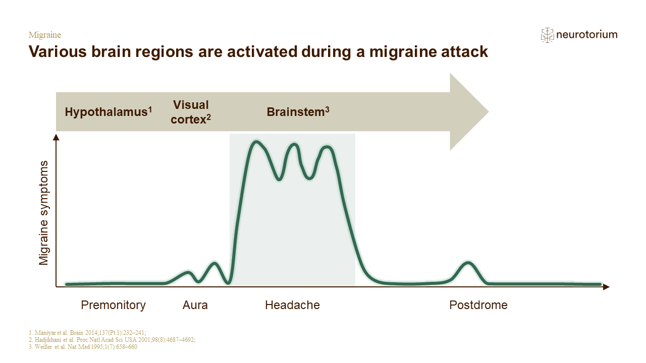
Several lines of evidence suggest activation of different regions of the brain at different stages of a migraine attack.
- During the premonitory phase, positron emission tomography (PET) studies have shown activation of the hypothalamus in individuals with nitroglycerin-triggered migraine attacks.[Maniyar et al., 2014]
- During aura, magnetic resonance imaging (MRI) studies have shown activation of the visual cortex in individuals who experience migraine attacks during the aura stage.[Hadjikhani et al., 2001] Furthermore, the slow and contiguously progressive nature of blood oxygenation level-dependent (BOLD) signal activation has been linked to the cortical spreading depression phenomenon.[Hadjikhani et al., 2001]
- During the headache phase of the migraine attack, PET studies have shown increased blood flow in some cerebral regions of the brain as well as in the brainstem.[Weiller et al., 1995]