

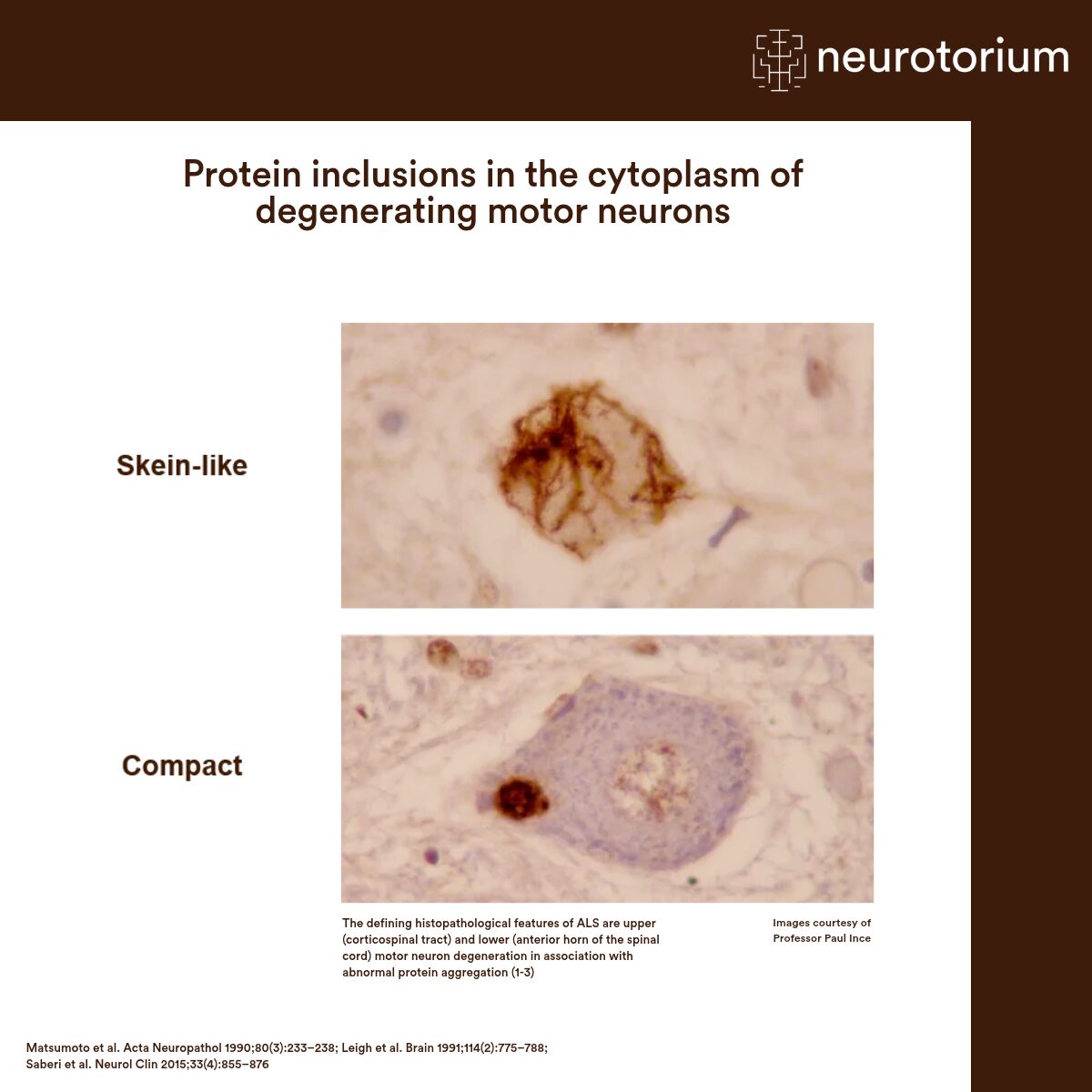
The defining histopathological features of ALS are upper (corticospinal tract) and lower (anterior horn of the spinal cord) motor neuron degeneration in association with abnormal protein aggregation.1-3 A complex array of neurodegenerative mechanisms leading to neuronal death are implicated in ALS, including glutamate excitotoxicity, oxidative stress, mitochondrial dysfunction, inflammation, and axonal transport disruption.4 TDP-43 pathology is thought to be central in ALS – hyperphosphorylated, ubiquitinated TDP-43 neuronal inclusions are observed in ~97% of cases.5
While there are currently numerous candidates under investigation, there are not yet currently any fluid or imaging biomarkers validated for routine clinical use6
Related content
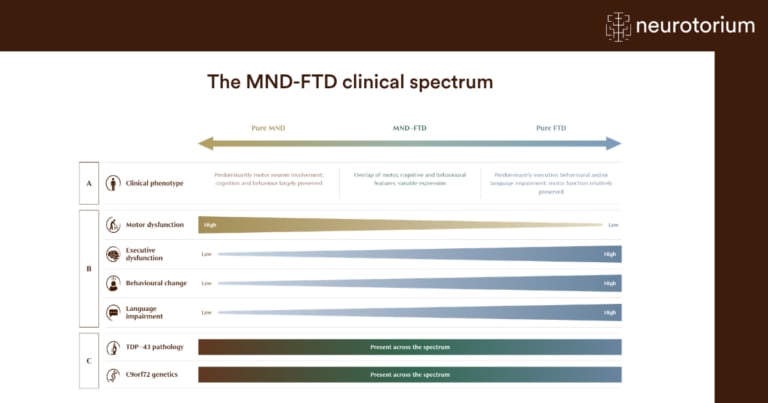
Motor neuron disease (MND) and frontotemporal dementia (FTD) were once seen as separate conditions, one affecting movement and the other cognition and behaviour. Growing evidence now places them on the same disease spectrum, linked by shared clinical features, genetic causes, and underlying pathology.
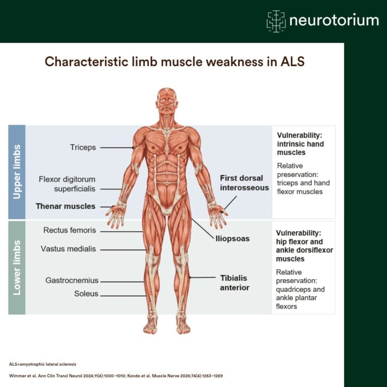
ALS is associated with characteristic patterns of muscle weakness, with selective vulnerability and relative preservation of specific upper- and lower-limb muscles.