Transcranial sonography (TCS) is one of the more promising imaging techniques as it is widely available and is more cost-effective than other methods, such as single-photon emission computed tomography (SPECT).5,6 In more than 90% of patients with PD, TCS reveals abnormally high-density tissue in the substantia nigra region of the brain, as compared with only 9% of healthy individuals.7 This finding appears to be stable during the course of the disease, and is thought to reflect increased amounts of iron.6,7 However, it does not necessarily reflect the degeneration of presynaptic dopaminergic nerve terminals – i.e., the rate of disease progression.7
References:
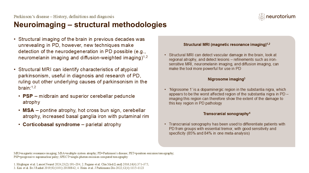
1.Höglinger GU, Adler CH, Berg D, et al. A biological classification of Parkinson’s disease: the SynNeurGe research diagnostic criteria. Lancet Neurol 2024; 23 (2): 191–204.
2.Pagano G, Niccolini F, Politis M. Imaging in Parkinson’s disease. Clin Med (Lond) 2016; 16 (4): 371–375.
3.Kim EY, Sung YH, Lee J. Nigrosome 1 imaging: technical considerations and clinical applications. Br J Radiol 2019; 92 (1101): 20180842.
4.Heim B, Peball M, Hammermeister J, et al. Differentiating Parkinson’s disease from essential tremor using transcranial sonography: a systematic review and meta-analysis. J Parkinsons Dis 2022; 12 (4): 1115–1123.
5.Schapira AHV. Recent developments in biomarkers in Parkinson disease. Curr Opin Neurol 2013; 26: 395–400.
6.Miller DB, O’Callaghan JP. Biomarkers of Parkinson’s disease: present and future. Metabolism 2015; 64 (3 Suppl 1): S40–S46.
7.Spiegel J, Hellwig D, Möllers MO, et al. Transcranial sonography and [123I]FP-CIT SPECT disclose complementary aspects of Parkinson’s disease. Brain 2006; 129 (5): 1188–1193.