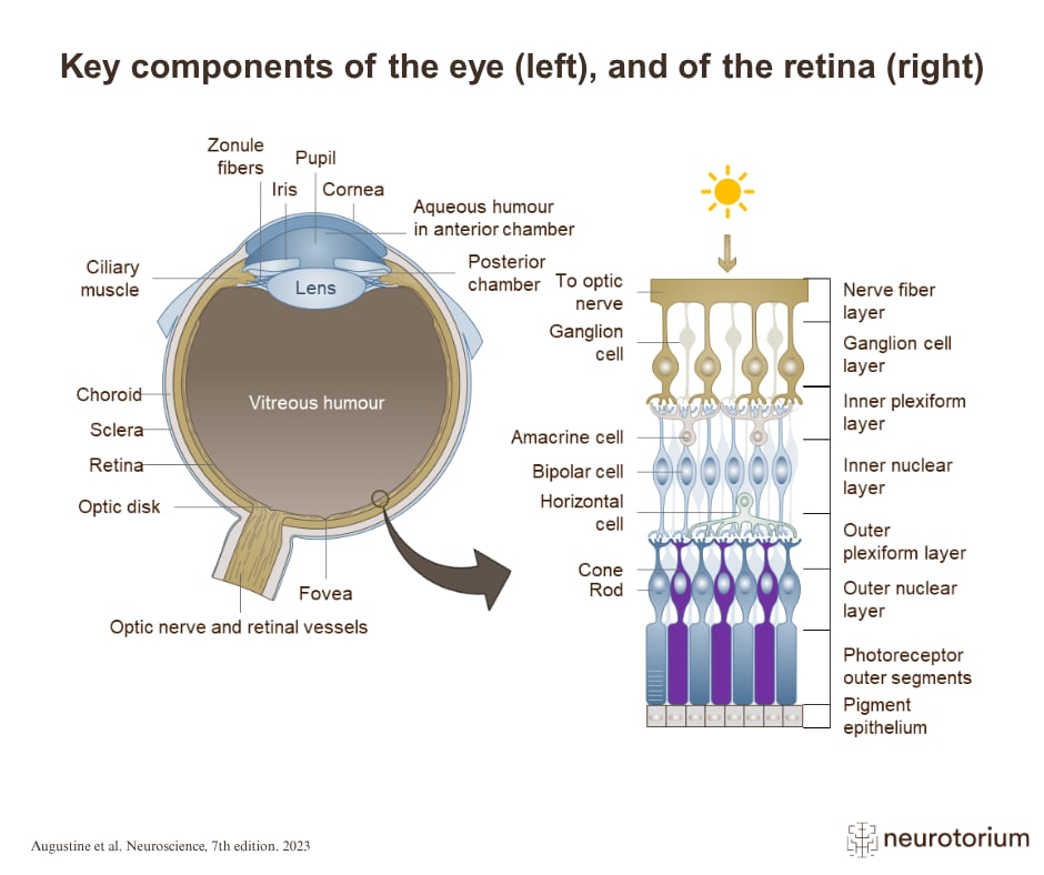
The retina is a thick (few hundred micrometres) sheet of neurons.2 It contains five major cell types, arranged in three cellular layers (i. photoreceptors [rods or cones]; ii. horizontal, bipolar and amacrine cells; iii. ganglion cells), separated by the outer and inner (synaptic) plexiform layers.2 The photoreceptor cells absorb light, convert it into a neural signal through a process called phototransduction, and pass the signal synaptically to bipolar cells, which in turn pass it to retinal ganglion cells in the innermost layer of the retina. The axons of the retinal ganglion cells form the optic nerve.2 The horizontal and amacrine cells provide the lateral connections in the outer and inner synaptic layers.2
Related content
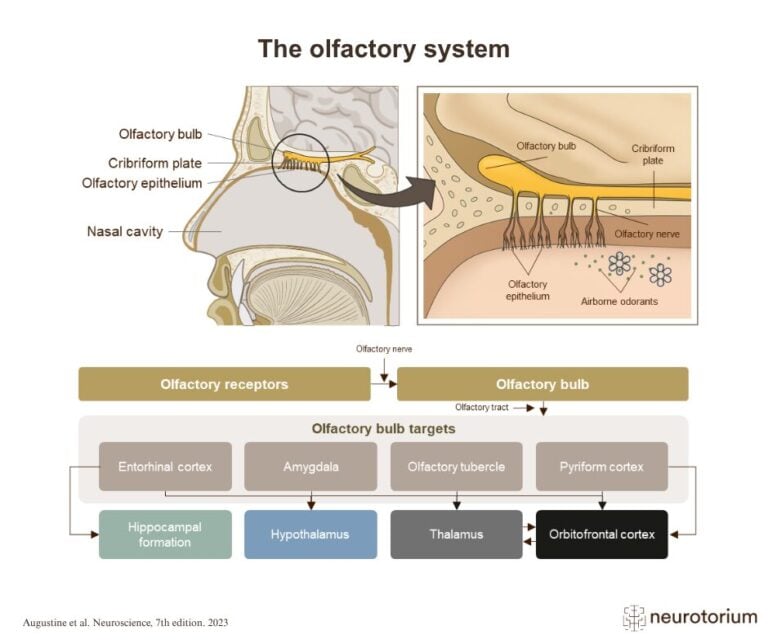
The olfactory system detects airborne odor molecules in the nasal cavity and transmits this information to the olfactory bulb. From there, signals are relayed to several brain regions involved in smell perception, memory, emotion, and behaviour.
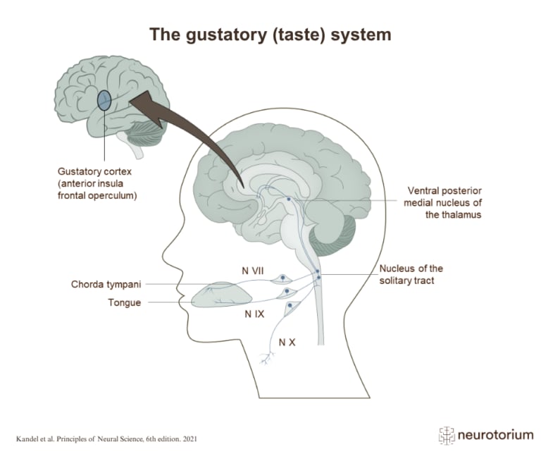
The gustatory system detects taste stimuli on the tongue and relays this information through brainstem and thalamic pathways to the gustatory cortex.
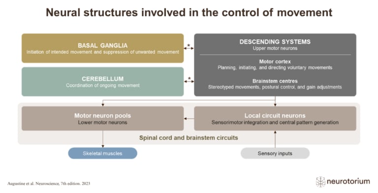
Movement is controlled by a network of brain and spinal cord structures that work together to plan, initiate, coordinate, and execute actions.