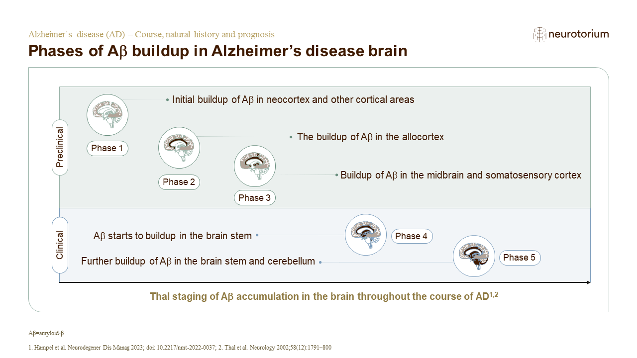
In vivo neuroimaging data from neuropathological studies show a spatiotemporal development of Aβ deposits in the brain, which originates in the cerebral regions and spreads from the neocortex, to the allocortex, to the brainstem, finally reaching the cerebellum.1,2 Aβ aggregation involves the formation of monomers and oligomers, which then develop into Aβ fibrils and plaques.3 Aβ40 and Aβ42 are the main components of Aβ aggregation, however Aβ42 aggregates into fibrils faster than Aβ40.3 Aβ accumulation in various brain regions results in a cascade of brain damage including abnormal tau phosphorylation, and dysfunction in neuronal physiology (i.e., oxidative stress, mitochondrial dysfunction, neuroinflammation, and impaired brain plasticity).3
Phases of Aβ buildup in Alzheimer’s disease brain