



Alzheimer’s disease (AD) is a progressive neurodegenerative disease characterized by cognitive and functional decline, including brain accumulation of b-amyloid and tau proteins. Since clinical presentations overlap between different diseases causing cognitive decline, an accurate diagnostic work-up of AD needs to demonstrate presence of the core pathologies, to minimize the risk of misdiagnosis. Biomarker research in this area over several decades has transformed clinical management of AD, with biomarkers increasingly available to reflect different aspects of brain pathophysiology relevant for AD. Recently, two major developments have had a particularly strong impact. The first is related to advances in minimally invasive technologies. We can now measure tau phosphorylated at threonine 217 (p-tau217) in blood as an indicator of AD pathology. This has turned biomarker testing from a procedure requiring a lumbar puncture or a PET camera into a simple blood draw, which allows molecular diagnostics at scale and in health care settings where it was previously not available. The second important development is relevant in several (but not all health contexts, depending on regulatory decisions) and is related to advances in treatments1,2. Anti-amyloid antibodies are now approved for use in many health care systems and have been shown to slow cognitive decline in early symptomatic AD. This means that a biomarker result can lead directly to a treatment decision rather than only to a diagnostic label.
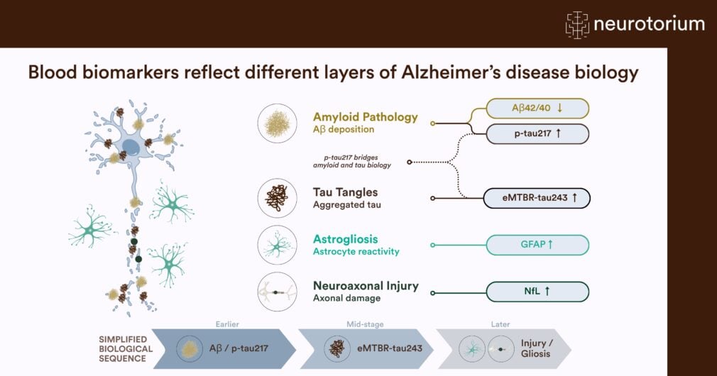
Figure 1. Overview of biomarkers in relation to cell types and pathologies
Blood biomarkers have been developed for the key pathological processes in Alzheimer’s disease. The biomarkers develop in an overall predictable fashion during the time course of the disease, in parallel with build-up of different brain changes.
The scale of the first shift, the rise of blood biomarkers (BBMs), is easy to underestimate. Over the last years there has been a surge in regulatory-approved BBMs for AD, largely based on p-tau217 with or without integration of b-amyloid related peptides. But one important challenge remains for clinical use of AD biomarkers. What clinicians now increasingly need to know is not just if the patient in front of them has AD pathology, but whether the pathology they have detected is severe enough to explain the symptoms at hand, how far along the disease course that patient is, and when imaging or CSF tests are still worth doing. This article summarizes where fluid biomarkers stand on those questions, with an emphasis on clinical application, and briefly on their use in research and clinical trials.
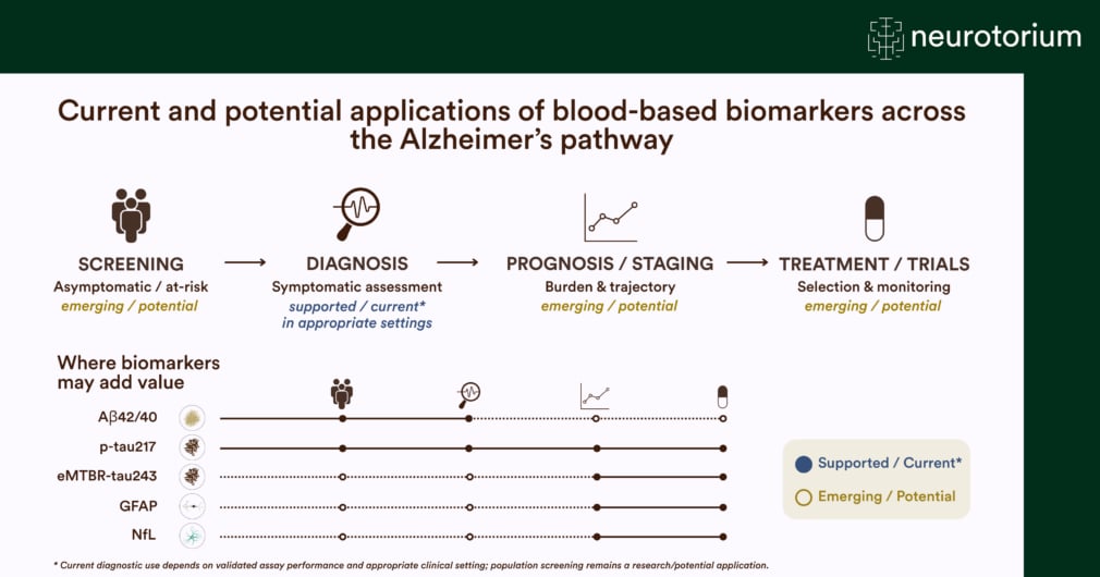
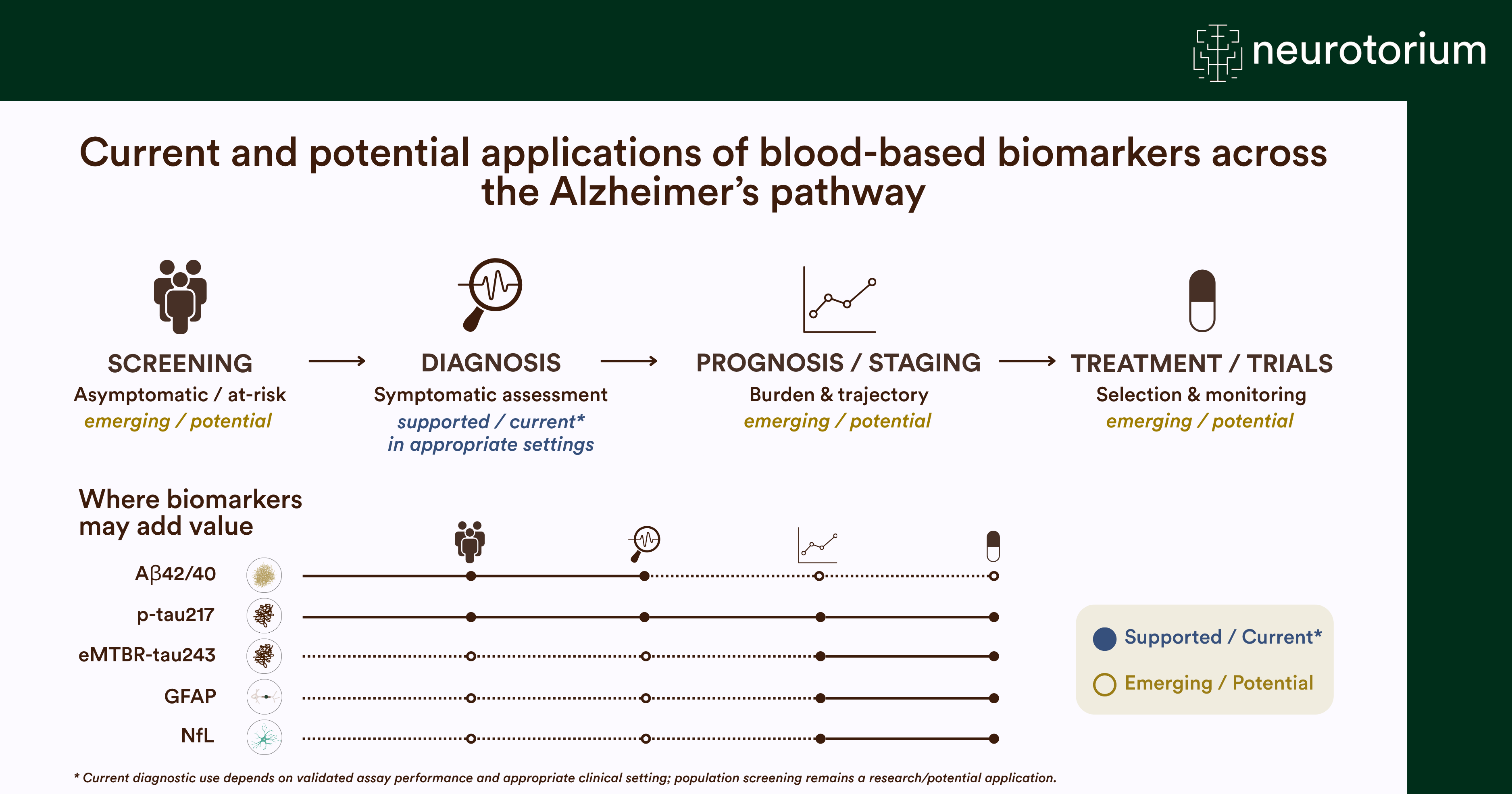
Figure 2. Current and potential applications for fluid biomarkers
Different blood biomarkers may fit different purposes in a health care system that integrates screening, diagnosis, prognostication, and treatment monitoring. Different blood biomarkers are already validated for some of these purposes, while others remain emerging or potentially useful.
A useful distinction: Core 1 and Core 2 biomarkers
The 2024 revised criteria for the diagnosis and staging of AD introduced a distinction that is now central to how fluid biomarkers are used3. Core 1 biomarkers become abnormal early and indicate that AD pathology is present. These biomarkers include e.g., CSF Aβ42/Aβ40, CSF or plasma p-tau217, and amyloid PET. They answer a binary question, namely “does this person have AD pathology?” Core 2 biomarkers become abnormal later and track the accumulation of tau tangles, which is the process most closely tied to symptoms. The archetypical biomarker in this class is tau PET, with emerging microtubule-binding region (MTBR) tau fragments being the most promising representative fluid biomarker (Table 1).
The distinction between core 1 and core 2 biomarkers matters because the two classes support different decisions. A positive core 1 result establishes that the disease process is underway, but it does not establish causation with cognitive symptoms, since amyloid pathology is present in a large proportion of cognitively normal people, especially over age 70. This is why we identify core 1 positivity in healthy control populations, without any significant cognitive impairment. In a memory clinic setting, a substantial proportion of core 1-positive patients have symptoms driven by something else than AD, e.g., cerebrovascular disease, Lewy body pathology, depression, sleep disorders, or a combination of factors. In those, AD may still be present in a very early stage (and be identified by the core 1 biomarkers) but not necessarily causing or even contributing to symptoms in a substantial way. In contrast, a positive core 2 result speaks to progressive AD pathology severity and can help clarify if AD is the likely cause of the symptoms or not.
| Biomarker | Class | What it indicates | Main clinical use | Key limitation |
| Plasma p-tau217 | Core 1 | Aβ pathology present | First-line test to detect or rule out AD pathology in patients with cognitive symptoms | Grey zone with a single cut-off; lower PPV in low-prevalence settings; reduced accuracy above age 80 |
| Plasma Aβ42/Aβ40 | Core 1 | Aβ pathology present | Reduces the intermediate range when combined with p-tau217 | Small dynamic range; adds little to p-tau217 accuracy alone |
| CSF Aβ42/Aβ40 or Aβ42/p-tau | Core 1 | Aβ pathology present | Confirmatory test; reference standard in many centres | Requires lumbar puncture |
| Plasma / CSF (e)MTBR-tau243 | Core 2 | Tau tangle pathology and its severity | Second step in p-tau217-positive patients, to judge whether AD explains symptoms and how advanced tangle pathology is | Mass spectrometry required; not yet widely available |
| CSF %p-tau205 | Core 2 | Tau pathology around tau PET positivity | Staging within a research panel | Not established in routine care |
| CSF α-synuclein SAA | Co-pathology | Lewy-related pathology | Explaining atypical presentations and discordant results | Requires lumbar puncture; no validated blood equivalent |
| Amyloid PET | Core 1 (imaging) | Aβ load, quantitatively | Confirmatory test in the grey zone; measuring plaque removal during treatment | Cost and availability |
| Tau PET | Core 2 (imaging) | Regional tangle burden | Prognosis; trial outcome; regional staging | Cost and availability |
Table 1. Fluid and imaging biomarkers for Alzheimer’s disease in clinical use
The 2024 revised criteria for the diagnosis and staging of AD introduced a distinction that is now central to how fluid biomarkers are used3. Core 1 biomarkers become abnormal early and indicate that AD pathology is present. These biomarkers include e.g., CSF Aβ42/Aβ40, CSF or plasma p-tau217, and amyloid PET. They answer a binary question, namely “does this person have AD pathology?” Core 2 biomarkers become abnormal later and track the accumulation of tau tangles, which is the process most closely tied to symptoms.
Core 1 and Core 2 follow the 2024 revised criteria3.
It is widely known that two patients with a positive standard AD biomarker test (e.g., CSF Aβ42/P-tau) may have grossly different clinical presentations, ranging from mild subjective cognitive impairment to severe dementia. The distinction between core 1 and core 2 biomarkers helps to resolve this common clinical conundrum of heterogeneity in disease severity and disease progression among patients who have positive results on common AD biomarkers.
Plasma p-tau217: accurate enough for routine care
Plasma p-tau217 is the best-performing single blood biomarker for AD pathology4, and the evidence base is now large enough to support use also outside specialist centres. In a prospective study of patients evaluated for cognitive symptoms in Sweden, a mass spectrometry-based test measuring the ratio of phosphorylated to non-phosphorylated tau217 (expressed as %p-tau217), alone or combined with the plasma Aβ42/Aβ40 ratio, identified AD pathology with a diagnostic accuracy around 90%. The comparison with usual care was striking. Primary care physicians reached 61% accuracy for clinical AD after examination, cognitive testing and a CT scan, and dementia specialists 73%, whereas the blood test reached 91% in both settings. Adding the Aβ42/Aβ40 ratio to %p-tau217 did not improve accuracy5. A subsequent study extended this to a fully automated, commercially available immunoassay in participants across four secondary care cohorts in Sweden, Spain and Italy plus a Swedish primary care cohort6. The test detected AD pathology with AUCs around 0.95.
A single threshold forces a trade-off between sensitivity and specificity. Using two cut-offs, a lower one to rule out and a higher one to rule in, raised the accuracy to 92-94% in both primary and secondary care. The cost of this procedure is that a proportion of patients (~15% in this study;6) are left in an intermediate or “grey zone” range that requires further testing.
Moving from “is AD present?” to “is AD causing the symptoms?”
If plasma p-tau217 tells us that amyloid pathology is present, it says considerably less about whether that pathology explains the patient’s complaints. This is where the MTBR-tau fragments have changed the playing field. MTBR-tau species were first identified to be abundant in AD brains. This led to development of assays for CSF MTBR-tau243, which turned out to be a specific marker of tau tangle pathology, rising in parallel with insoluble tau aggregates in a way that phosphorylated tau species do not7. More recently, MTBR-tau243 has also been measured in plasma as the endogenously cleaved fragment eMTBR-tau243, which retains the key properties of the CSF version8.
If plasma p-tau217 tells us that amyloid pathology is present, it says considerably less about whether that pathology explains the patient’s complaints. This is where the MTBR-tau fragments have changed the playing field
In a prospective study of patients with cognitive symptoms, we tested a two-step strategy. Of 572 patients, 61% were positive for plasma %p-tau217, and of those, 97% were amyloid-positive by CSF or PET9. This confirmed that plasma p-tau217 was an excellent test of amyloid pathology. But only 57% had established AD, meaning both AD-specific pathology and a clinical picture consistent with it. In other words, a positive p-tau217 result alone had a positive predictive value of only 57% for symptomatic AD in this memory clinic sample. Adding plasma eMTBR-tau243 as a second step changed that substantially. Among the p-tau217-positive patients, 55% were also eMTBR-tau243 positive, and this identified established AD with 81% accuracy, a positive predictive value of 84% and a negative predictive value of 77%. Positive eMTBR-tau243 also predicted faster subsequent cognitive decline and faster tau tangle accumulation as measured by PET. The eMTBR-tau243 test could also be used to stratify tau PET burden and identified a high tau PET load with 87% accuracy.
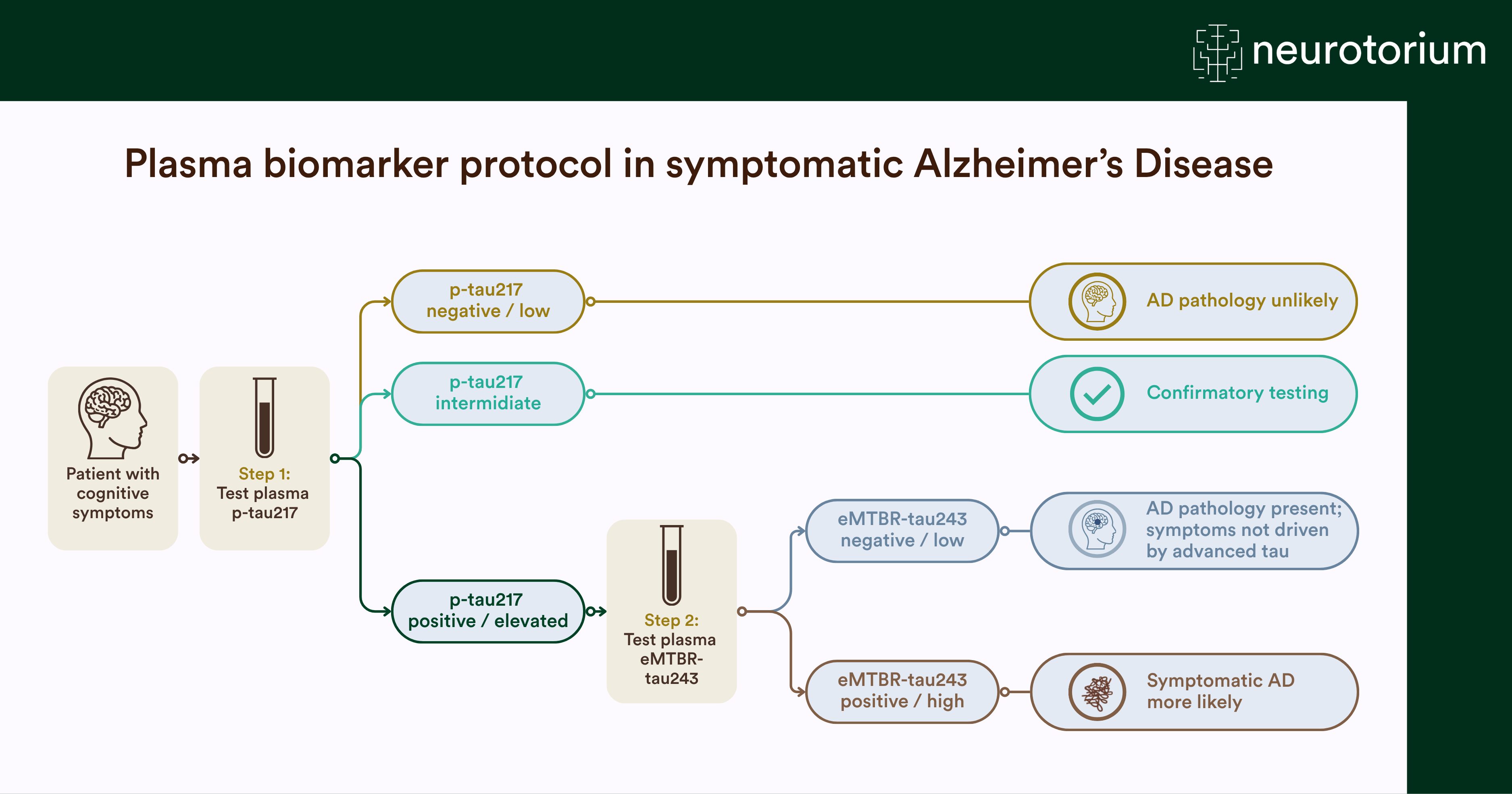
The clinical logic is appealing in its simplicity. We have imagined a protocol where clinicians first test with plasma p-tau217 to establish whether amyloid pathology is present, and then test plasma eMTBR-tau243 in those who are p-tau217 positive to judge the likelihood that tau pathology has advanced far enough to be a likely cause or significant contributor to the symptoms (Figure 1). However, currently available plasma assays cannot unambiguously resolve patient status. Gray-zone patients will remain, requiring secondary tests or intensified work-up. There may also be sensitivity issues with plasma eMTBR-tau243, so that some patients may present with falsely low levels of the biomarkers. The biomarkers still therefore need to be interpreted carefully in relation to clinical symptoms, possible differential diagnoses, and auxiliary findings.
When does each biomarker change?
Interpreting a biomarker result requires knowing where in the disease course it becomes abnormal (Figure 3). Using mass spectrometry measurements of plasma and CSF tau species, with disease duration estimated from amyloid and tau PET, the ordering is now reasonably clear. Sensitive p-tau217 measures changes earliest, rising just before amyloid PET becomes positive. Other phosphorylated tau species change later, with smaller dynamic ranges and earlier ceiling effects. (e)MTBR-tau243 and p-tau205 (another phosphorylation variant of tau) change around the time tau PET becomes positive, and (e)MTBR-tau243 continues to track cortical tau burden into later stages, where the phosphorylated tau markers have plateaued10,11. This staggered sequence is what may make a biomarker panel more informative than any single marker. When measured together, plasma p-tau217, p-tau205 and MTBR-tau243 report on different biological events rather than three versions of the same one12.
Interpreting a biomarker result requires knowing where in the disease course it becomes abnormal. Using mass spectrometry measurements of plasma and CSF tau species, with disease duration estimated from amyloid and tau PET, the ordering is now reasonably clear.
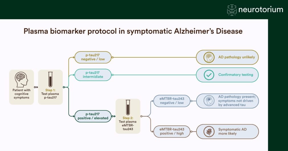
Figure 3. A two-step blood-based algorithm
A workflow has been suggested to integrate early Alzheimer biomarkers (p-tau217, a core 1 biomarker) for initial assessment of presence of Alzheimer pathology. Patients may undergo further testing, e.g., with advanced tests (CSF or PET) in selected cases with intermediate p-tau217 levels. Among those with elevated p-tau217 levels, eMTBR-tau243 may be assessed to determine likelihood for presence of significant tau pathology, supporting that Alzheimer pathology contributes substantially to cognitive decline.
Where PET still matters
One potential conclusion from the accuracy figures above would be that blood tests can simply replace amyloid PET or CSF testing. However, this conclusion may be premature, for three specific reasons13. First, the issue of the grey zone. As stand-alone tests, blood biomarkers show up to 90% sensitivity and specificity relative to amyloid PET. Two-threshold strategies improve on that, but by design they leave an intermediate group, where roughly 10-40% of individuals (depending on the setting and the assay) are still in need of a confirmatory test with PET or CSF to resolve their pathology status. Second, the issue of early disease. In cognitively unimpaired individuals, or patients with very mild cognitive signs, the prevalence of AD pathology is lower, which naturally reduces the positive predictive value (as explained above) of any test and enlarges the grey zone. This early-stage population is particularly relevant for very early intervention trials, which makes the reduced performance of BBMs in this context even more problematic. Third, and most fundamentally, blood biomarkers do not measure how much amyloid is in the brain. They are good at classifying amyloid status but cannot reliably estimate the quantitative amyloid load14. They also do not track plaque removal after immunotherapy. If the question is whether a treatment is working (whether plaques are being cleared), amyloid PET currently has no fluid substitute. A reasonable division of labour is therefore emerging. We can imagine BBMs as the first-line test for triage and for ruling out AD pathology, with PET (or CSF) as a confirmatory test in intermediate cases and in early-stage populations, and PET as the measure of treatment response (Figure 2).
What cerebrospinal fluid is still needed for
The rise of blood tests has not made CSF obsolete, but it has narrowed and sharpened its role. CSF remains the more informative fluid when the question is broader than “is this AD?” Co-pathology is the clearest example. There are now α-synuclein seed amplification assays that can detect Lewy-related pathology in CSF. Although used mainly in research, these also start to be available clinically in advanced specialized settings. This pathology is common in older people and contributes independently to cognitive decline, beyond the AD changes15. In current clinical practice, three situations still argue for a lumbar puncture (when PET is not available, too expensive or cumbersome). The first is when there is an intermediate blood result that must be resolved. The second is when a blood result conflicts with the clinical picture. The third is when the presentation is atypical enough that non-AD pathologies (e.g., α-synuclein) need to be assessed directly. Outside those situations, blood testing is rapidly becoming the reasonable first choice for most patients with cognitive symptoms.
Use in research and clinical trials
The impact of fluid biomarkers on trials has been at least as large as on clinical care. Screening for amyloid positivity with PET or CSF is slow and expensive. Plasma p-tau217 allows a large population to be pre-screened cheaply so that confirmatory testing is performed mainly in those likely to be eligible, which reduces screen failures substantially. This matters most for the trials that are hardest to fill. Prevention trials recruit cognitively unimpaired people, in whom the prevalence of AD pathology is low and the number who must be approached to enroll one eligible participant is large. A cheap first-line blood test is what makes those trials arithmetically feasible, even when its positive predictive value in that population is modest and confirmatory imaging or CSF is still needed. Beyond selection, the next step is for accessible fluid markers to become outcome and target-engagement measures. Because MTBR-tau243 reflects tau aggregation specifically, it can potentially show whether a tau-directed drug is doing what it is meant to do. We are now living in an era of emerging therapies engaging not only amyloid but also tau pathology in AD. It will be fascinating to learn if such treatments can engage tau pathology in a meaningful way, and if that can be mirrored by relevant changes in MTBR-tau243 concentrations. Plasma biomarkers are also extending AD research into populations where imaging or CSF is difficult, including in countries or health care settings with limited resources.
We are now living in an era of emerging therapies engaging not only amyloid but also tau pathology in AD. It will be fascinating to learn if such treatments can engage tau pathology in a meaningful way, and if that can be mirrored by relevant changes in MTBR-tau243 concentrations
Caveats before ordering a blood test
Several practical points determine whether a blood biomarker result can be trusted, or provide clinical value. At present, clinical use should focus on symptomatic individuals, with blood biomarker results interpreted within specialist services. For asymptomatic individuals, a positive biomarker result does not mean that symptomatic AD is inevitable; its clinical implications remain uncertain and require further research. For example, the assays are not interchangeable. Cut-offs derived for one platform do not transfer to another, and mass spectrometry-based and immunoassay-based measures of p-tau217 do not perform identically in every setting. For example, in primary care, in the oldest patients, and in the earliest disease stages, the mass spectrometry measure has performed better. A result must always be interpreted against the cut-off validated for that specific assay. Furthermore, setting changes performance. We have seen that even the same test yields different predictive values in a memory clinic and in primary care, and lower predictive values in cognitively unimpaired populations. Pre-analytical handling also matters. Tube type, plate format and sample processing can shift measured concentrations enough to move a patient across a fixed cut-off, which is why standardized protocols and thorough laboratory quality control are very important. Careful adjustment by valid reference protein measures may perhaps increase the robustness and value of already high-performing biomarkers 16. And finally, we must remember that a positive test is not a diagnosis. This is the most important caveat clinically, since amyloid pathology is common in older people, and many p-tau217-positive memory clinic patients have symptoms attributable to other causes than AD. A biomarker result should therefore still be clearly anchored with a clinical assessment, and not substitute it.
Outlook
The field has moved quickly from asking whether a blood test can detect AD to asking what a panel of blood tests can tell us about an individual’s disease stage, prognosis and likely treatment response. Plasma p-tau217 has largely settled the first question. Plasma eMTBR-tau243 is the most promising candidate for the second, and its combination with p-tau217 in a sequential algorithm is, in my view, the most clinically consequential development in fluid biomarkers since p-tau217 itself. What remains is the unglamorous work that determines whether any of this reaches patients. Researchers, industry and laboratories need to bring scalable assays to clinical practice. They need to harmonize those assays and cut-offs across platforms, and establish how results should be communicated, and define clearly when imaging or CSF is still required. In practice, specialist clinicians may increasingly integrate biomarker readouts with other relevant clinical information – supporting more precise approaches to diagnosis, prognosis, and treatment selection as new therapies emerge.
Related content
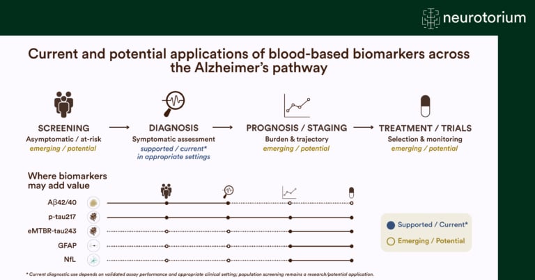
Overview of how blood-based biomarkers may support screening, diagnosis, staging, prognosis, and treatment decisions across the Alzheimer’s disease continuum.
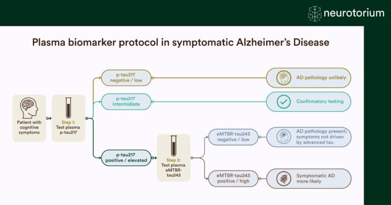
Overview of a plasma biomarker testing pathway for symptomatic Alzheimer’s disease, using p-tau217 and eMTBR-tau243 to guide diagnostic assessment and identify underlying pathology.
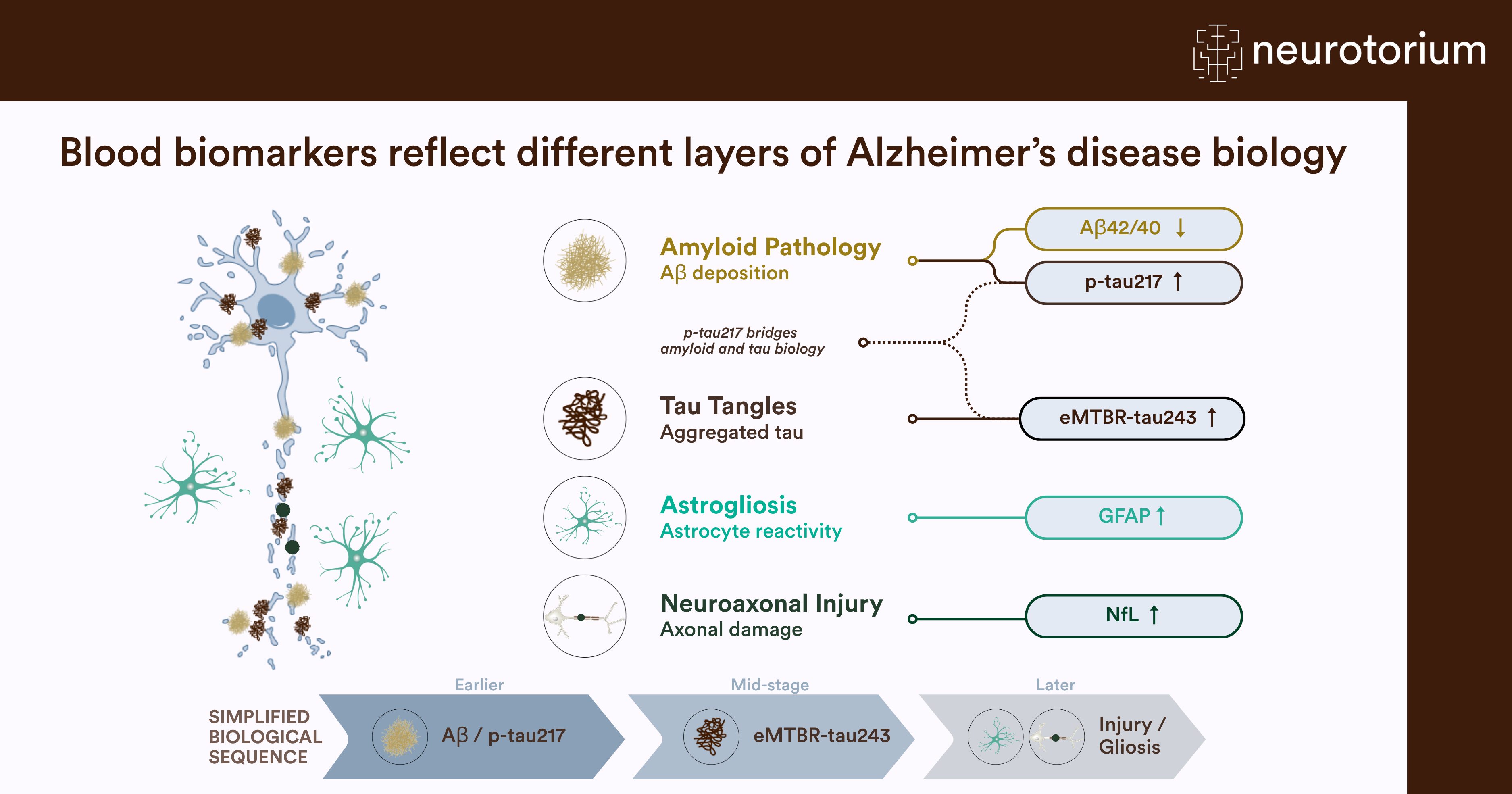
Overview of blood-based biomarkers that capture distinct aspects of Alzheimer’s disease pathology and progression.