
Images

Filter

search
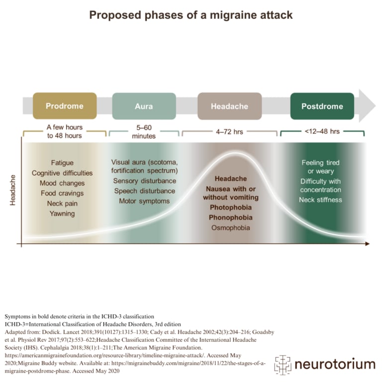
image Image Model showing the stages of a migraine attack and associated symptoms over time
Proposed phases of a migraine attack

Migraine attacks can be broken down into four phases: prodrome, aura, headache, and postdrome, as shown on the image. The exact timing of each phase is highly variable; the headache phase of a migraine can range from 4–72 hours in adults, and 2–48 hours in children.
29.08.2023 Migraine
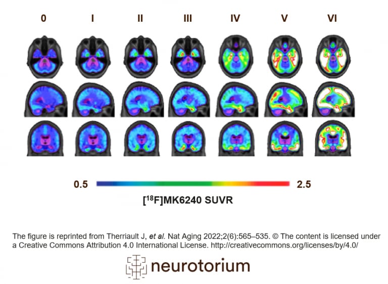
image Image In vivo quantification of tau can be performed using tau-PET (PET: positron emission tomography). Tau-PET binds to insoluble neurofibrillary tangles, another core pathological feature of AD.
PET tau across stages of Alzheimer’s disease

In vivo quantification of tau can be performed using tau-PET (PET: positron emission tomography). This illustration represents the stages of tau pathology measured by PET.
24.08.2023 Alzheimer’s Disease
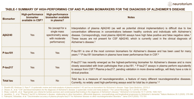
image Image Summary of high-performing CSF and plasma biomarkers for the diagnosis of Alzheimer’s disease (AD).
Summary of high-performing CSF and plasma biomarkers for the diagnosis of Alzheimer’s disease (AD)

Currently, CSF p-tau181, Aβ42/40, and total tau are used to diagnose AD in clinical practice. P-tau217 has recently gained attention due to its high sensitivity and specificity for AD.
24.08.2023 Alzheimer’s Disease
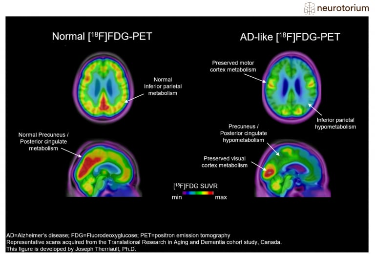
image Image Representative images of Fluorodeoxyglucose PET ([18F]FDG-PET) images in a control subject (left) and a patient with advanced Alzheimer’s Disease (AD) (right).
PET imaging of brain metabolism in Alzheimer’s disease

Representative figures of Fluorodeoxyglucose PET ([18F]FDG-PET) images in a control subject and a patient with advanced Alzheimer’s Disease (AD).
24.08.2023 Alzheimer’s Disease
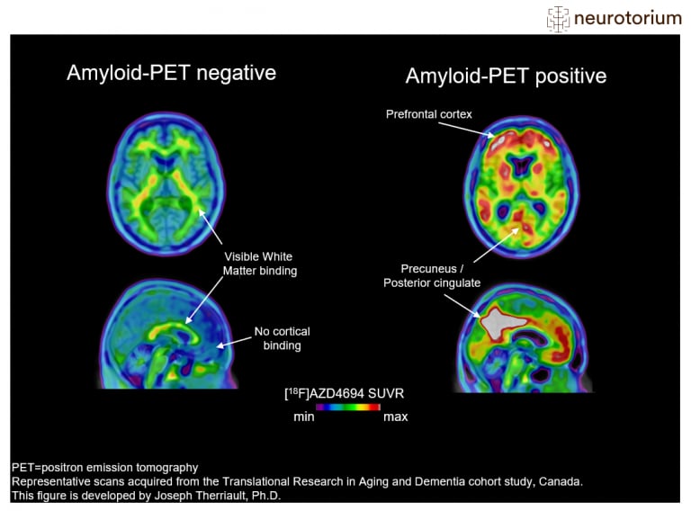
image Image Examples of amyloid-negative (left) and amyloid-positive (right) positron emission tomography (PET) scans.
Amyloid-PET imaging in Alzheimer’s disease

Examples of amyloid-negative and amyloid-positive positron emission tomography (PET) scans. In this figure, the amyloid-PET ligand [18F]AZD4694 is shown.
24.08.2023 Alzheimer’s Disease
image Image
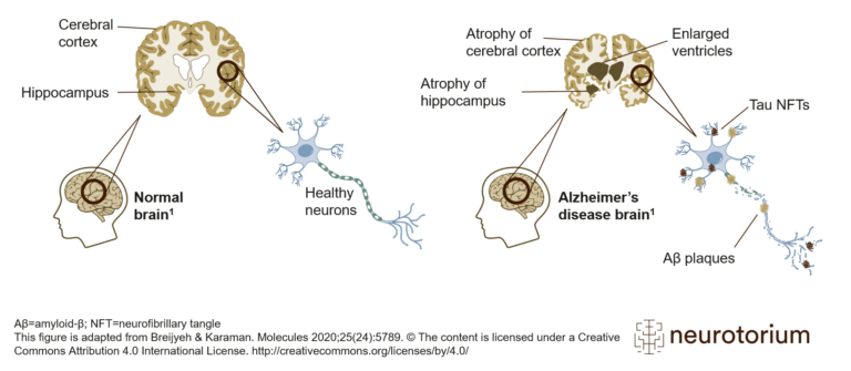
Alzheimer’s disease brain with amyloid plaque and neurofibillary tangles

Schematic representation of a healthy brain and Alzheimer’s disease (AD) brain. Compared to the healthy brain, the brain with AD, as the disease progresses, features an atrophied cerebral cortex, shrinkage of the hippocampus, and enlargement of the ventricles.

24.08.2023 Alzheimer’s Disease

No results matched your search.
Please try again.

task_alt