


Images
Filter
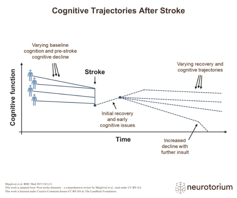
There are different paths of recovery that a patient can take after a stroke, which range from some degree of cognitive recovery to a steep decline and recurrent strokes.
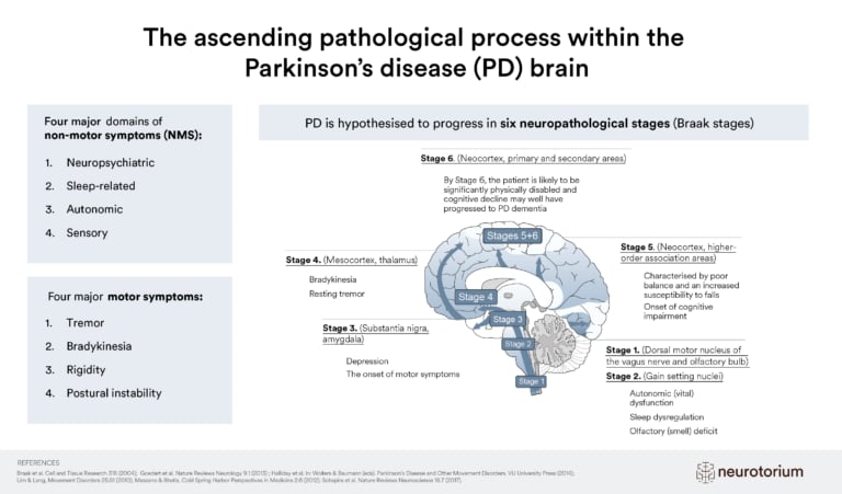
The Braak staging scheme is a valuable model for conceptualizing PD progression and categorizes PD into stages of neuropathological changes.
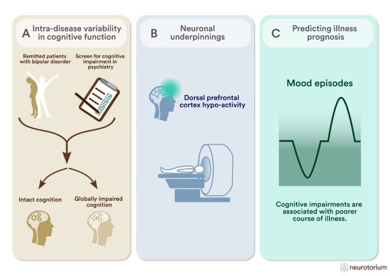
To study intra-disease variability in cognitive function, researchers employ data-driven clustering methods to differentiate patients with widespread cognitive abnormalities from patients who are cognitively intact.
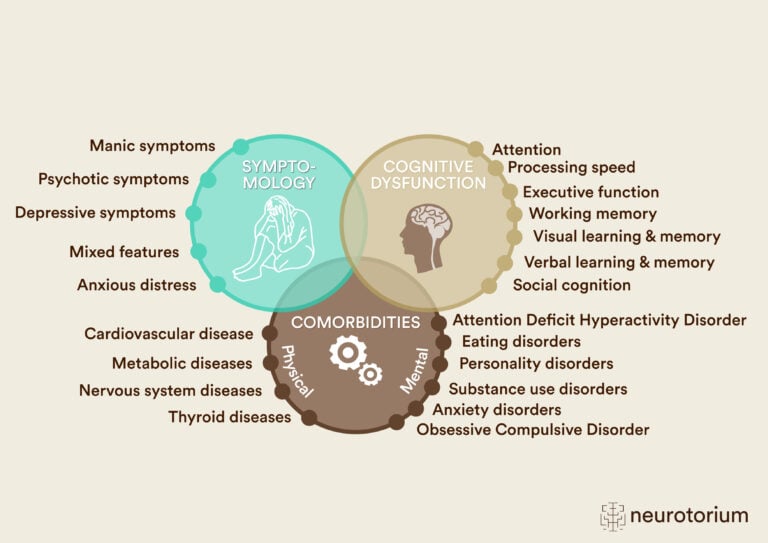
Bipolar disorder is associated with marked clinical and phenotypic heterogeneity, including differences in symptomatology, medical and psychiatric comorbidities, and neurocognitive function.
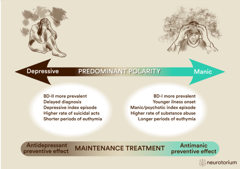
Depressive predominant polarity is typically defined as having at least two-thirds of lifetime episodes being depressive, whereas manic predominant polarity refers to at least two-thirds of prior episodes being (hypo)manic episodes.
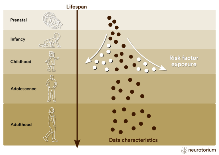
By integrating multiple data modalities this approach may yield information on risk architectures that can be useful for personalized psychiatry.
No results matched your search.
Please try again.