
Index for
slide deck
Regional Neuroscience
Regional Neuroscience
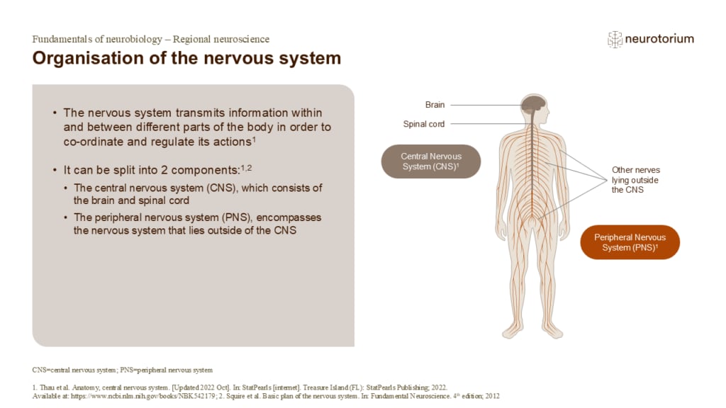
Organisation of the nervous system
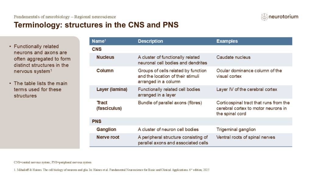
Terminology: structures in the CNS and PNS
The peripheral nervous system (PNS)
The peripheral nervous system (PNS)
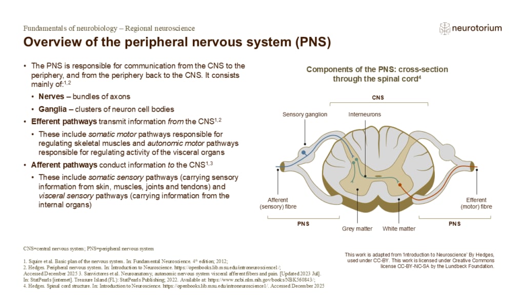
Overview of the peripheral nervous system (PNS)
The cell bodies of efferent pathways (motor and autonomic) lie in the spinal cord of the central nervous system (CNS) and peripheral nerves contain axons from these neuronal cell bodies as well as those situated in peripheral ganglia.1,2 The central/peripheral distinction…
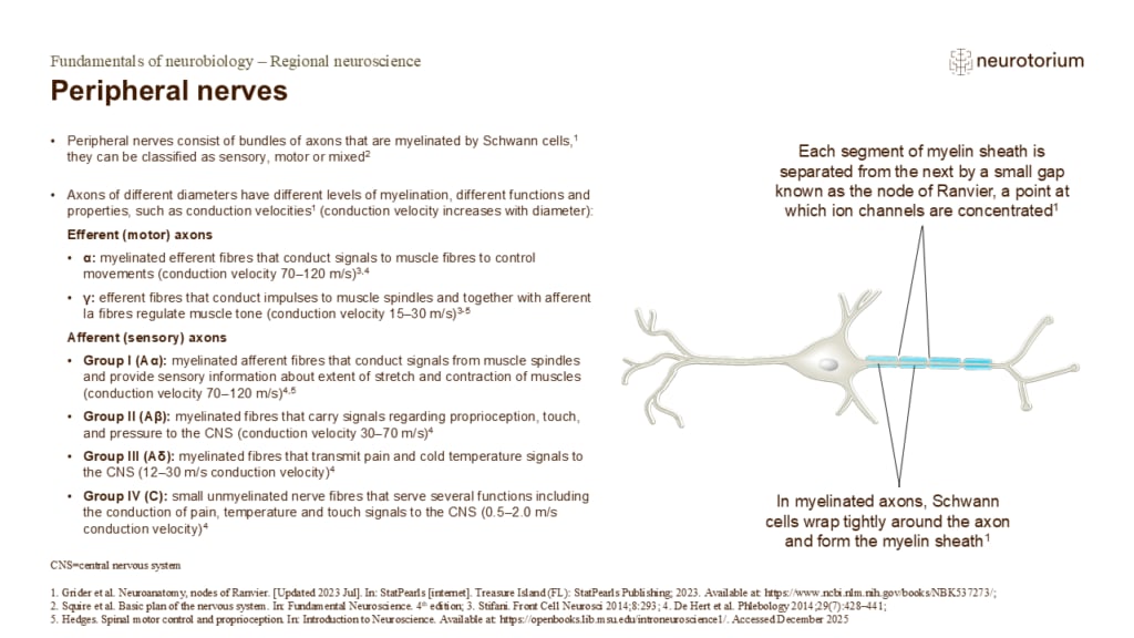
Peripheral nerves
Because of the electrical properties of the myelin sheath, and the concentration of sodium channels at nodes of Ranvier, conduction velocities in myelinated axons are much greater than those in unmyelinated axons.1
Clinical considerations: The diagnosis of peripheral neur…
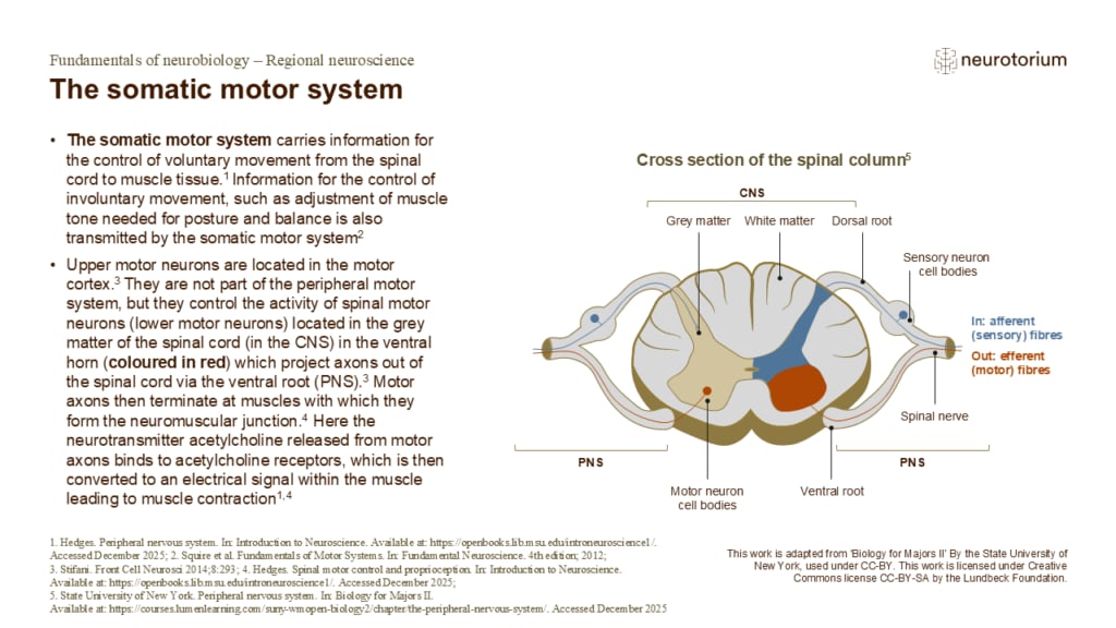
The somatic motor system
The cell bodies of large myelinated axons reside in the grey matter of the spinal cord in the anterior horn.6,7 They send their axons out of the ventral root and either directly into peripheral nerves, or nerve plexuses (a branching network of intersecting nerves) which g…
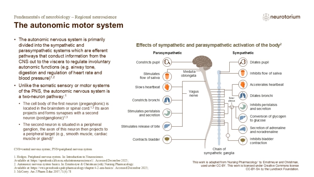
The autonomic motor system
The parasympathetic and sympathetic systems differ with respect to the neurotransmitters used: both use acetylcholine as a transmitter in the preganglionic neurons, but most postganglionic sympathetic neurons use noradrenaline whereas postganglionic parasympathetic neuron…
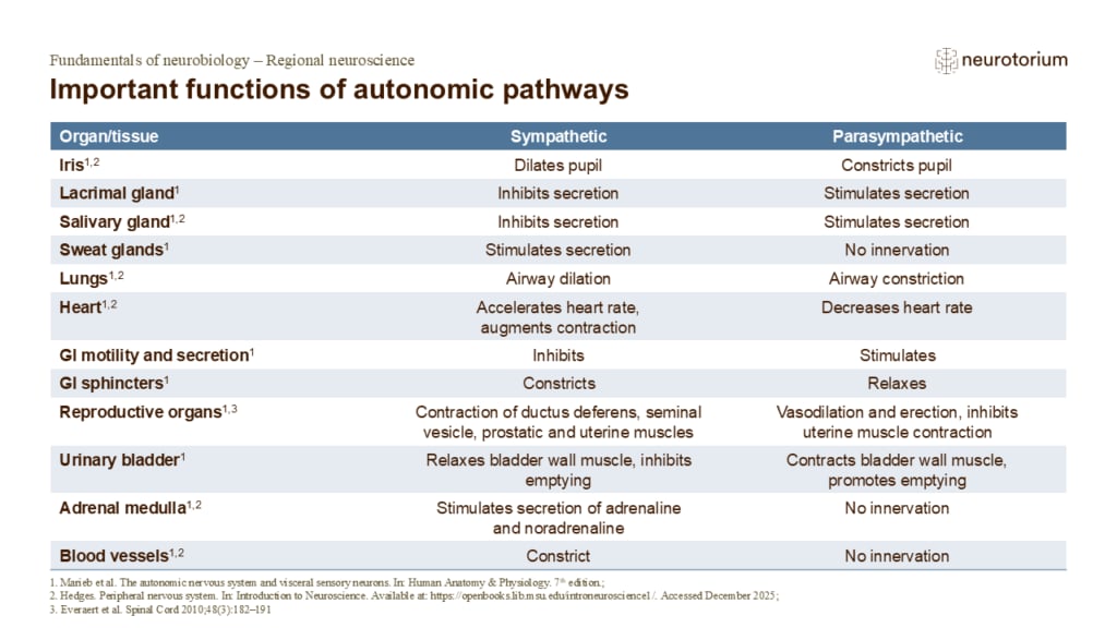
Important functions of autonomic pathways
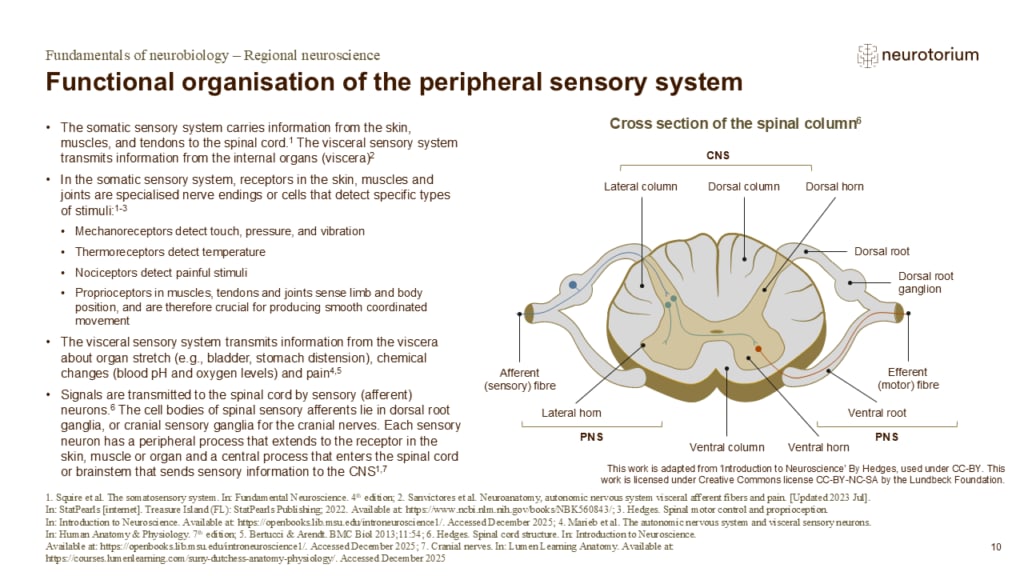
Functional organisation of the peripheral sensory system
Specialised sensory receptors in the periphery convert stimuli (e.g. touch, heat) into action potentials in a process known as sensory transduction.8
References:
1. The somatosensory system. In: Squire L, Berg D, Bloom FE, Lac Sd, Ghosh A, Spitzer NC. Fundamental Neurosci…
The central nervous system (CNS)
The central nervous system (CNS)
Regions of the central nervous system
References:
1. Thau L, Reddy V, Singh P. Anatomy, central nervous system. [updated October 2022]. In: StatPearls [internet]. Treasure Island (FL): StatPearls Publishing; 2022. Available at: https://www.ncbi.nlm.nih.gov/books/NBK542179/.
2. Shenoy SS, Lui F. Neuroanatomy, …
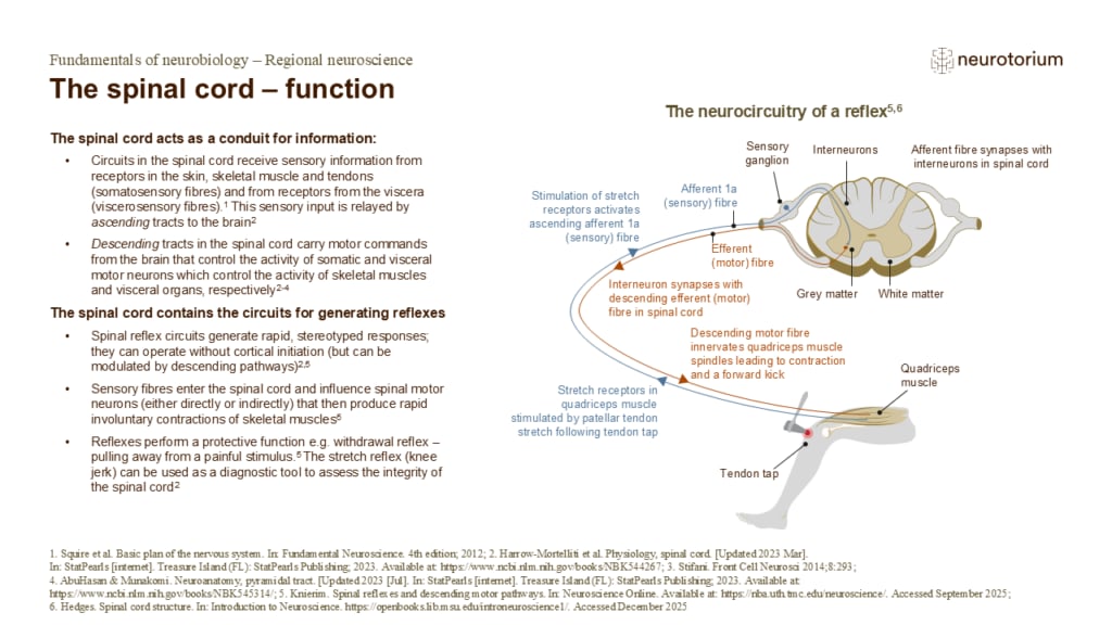
The spinal cord – function
Because so many important functions are concentrated into such a small region of the CNS, injury to the spinal cord can cause devastating neurological impairment.2 Damage to the thoracic spinal cord can produce paraplegia (paralysis of both legs), and loss of bowel and bl…
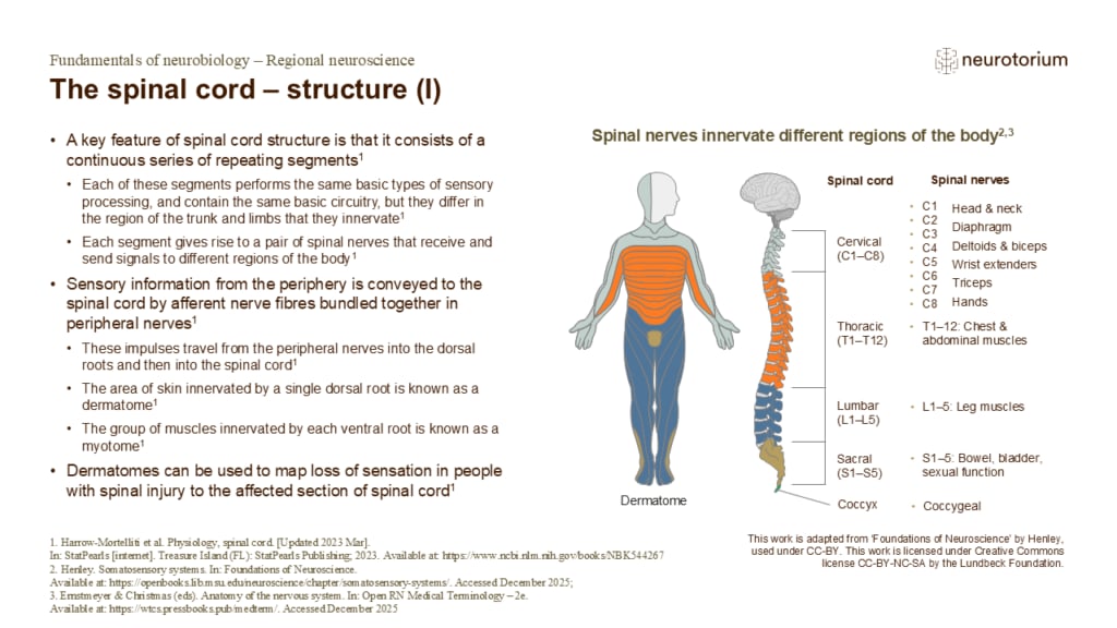
The spinal cord – structure (I)
There are 31 segments of the spinal cord grouped into regions:1,3,4
- 8 cervical (C1–C8): head, neck, arms, hands, and diaphragm
- 12 thoracic (T1–T12): chest and upper abdominal muscles
- 5 lumbar (L1–L5): lower back, hips, thighs, and knees
- 5 sacral (S1–S5): pelvic organs, l…
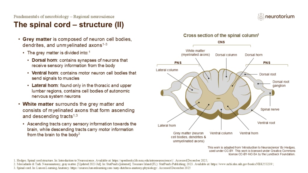
The spinal cord – structure (II)
The grey matter is located centrally in the spinal cord, forming a butterfly shape when viewed in cross section.4
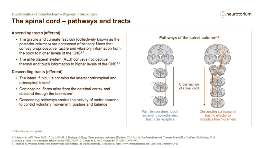
The spinal cord – pathways and tracts
Ascending tracts
The posterior (dorsal) spinocerebellar and anterior (ventral) spinocerebellar tracts are located on the lateral surface of the spinal cord and convey proprioceptive information to the cerebellum.5
Descending tracts
The anterior funiculus contains reticulo…
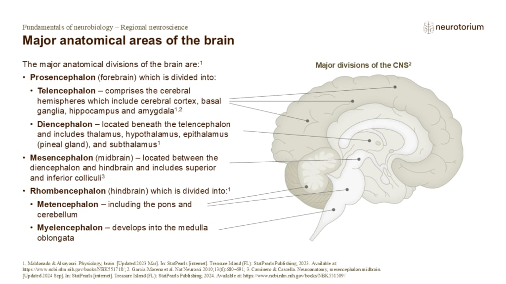
Major anatomical areas of the brain
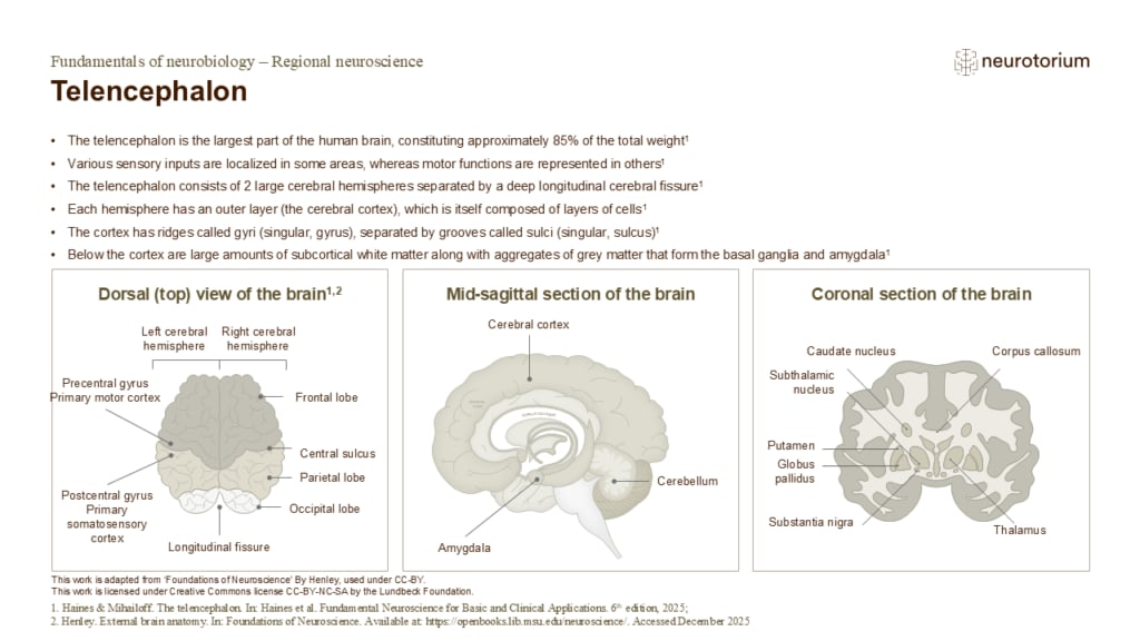
Telencephalon
Information flows into and out of the cerebral cortex via the subcortical white matter. The myelinated fibres forming the white matter are grouped into:
1. Association fibre bundles that connect gyri within one hemisphere
2. Commissural fibres that connect the two hemisph…
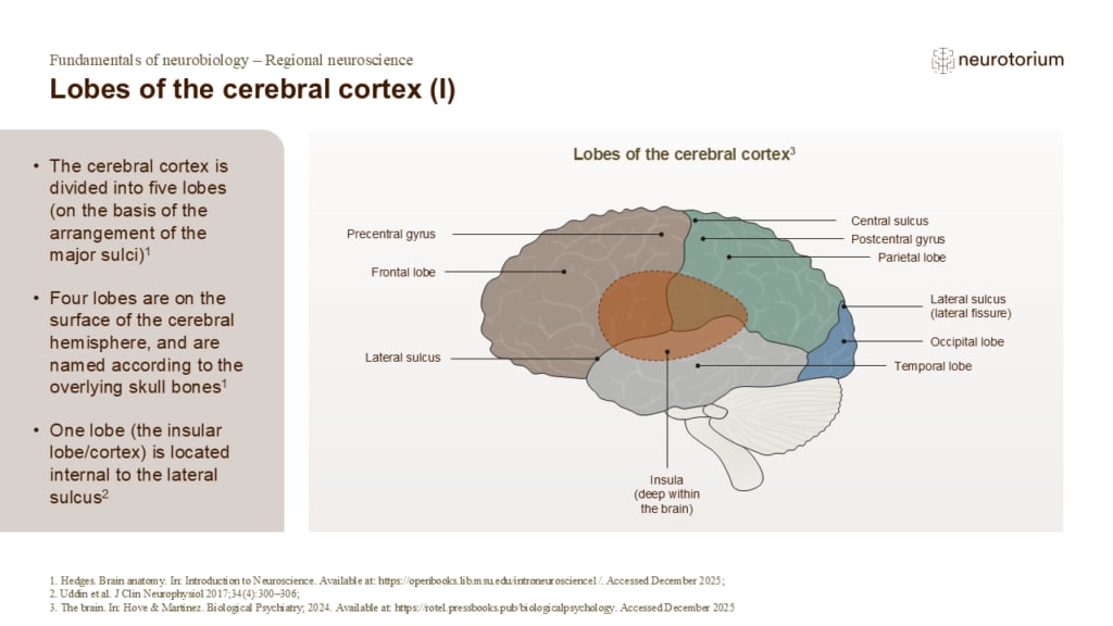
Lobes of the cerebral cortex (I)
Lobes of the cerebral cortex (II)
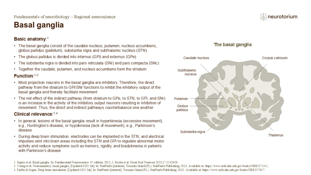
Basal ganglia
The basal ganglia are a group of subcortical nuclei that are important in motor control.1
Pathways:
- The caudate and putamen (striatum) the main input structures of the basal ganglia. GPi and SNr are the main output structures1,2
- Basal ganglia receives excitatory input fr…
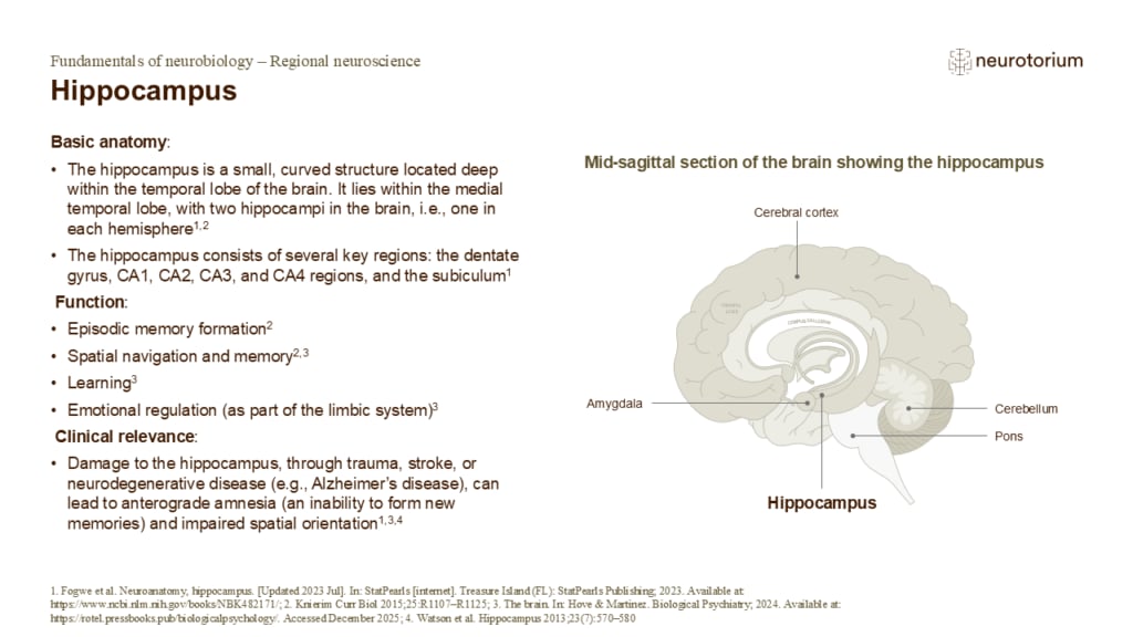
Hippocampus
Structure continued:
- The dentate gyrus is a key region that plays a role in forming new memories and in neurogenesis5
- The CA1 and CA3 regions are subfields responsible for the processing and relay of information, particularly in memory formation and retrieval6
- The subicu…
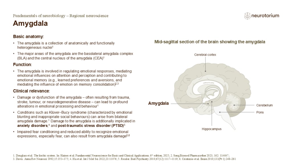
Amygdala
Structure continued:
- The BLA receives input from widespread cortical areas as well as from sensory nuclei of the thalamus.1,7 It therefore has access to high-level information from association areas as well as lower-level sensory information7
- The BLA projects to the CEA …

Thalamus
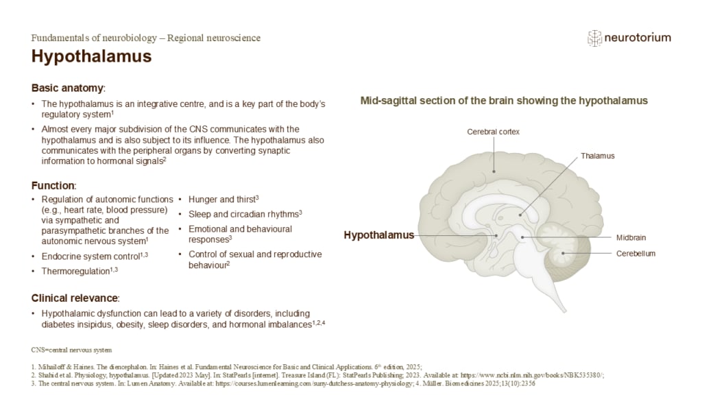
Hypothalamus
Regulation of autonomic functions: heart rate, blood pressure, and digestion are regulated by the hypothalamus, via its influence on the sympathetic and parasympathetic branches of the autonomic nervous system.1,3
Endocrine system control: The hypothalamus connects the ne…
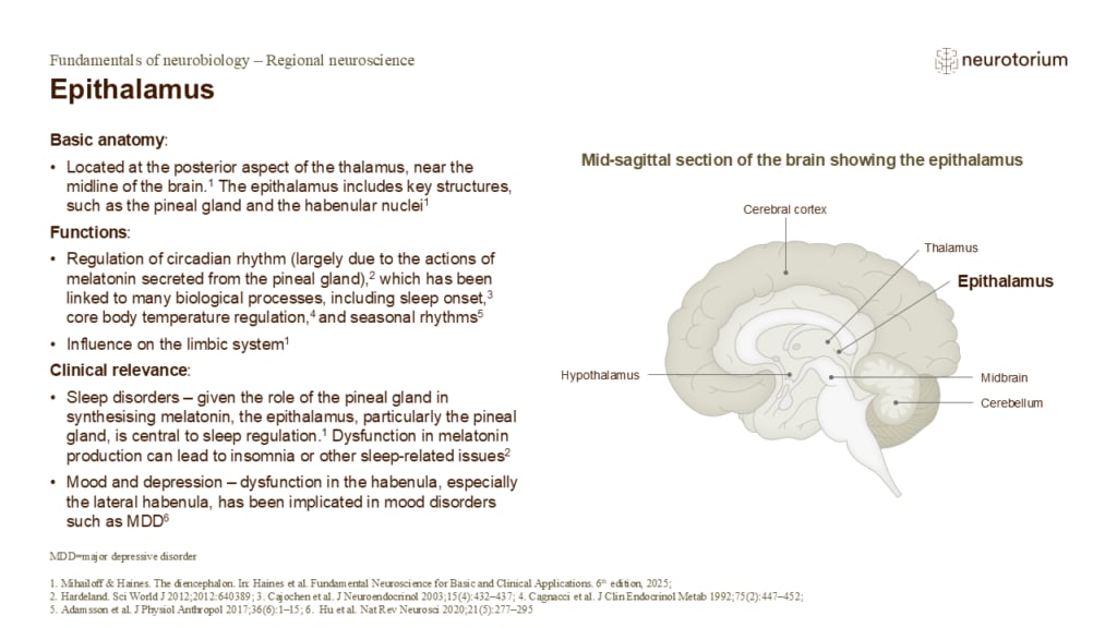
Epithalamus
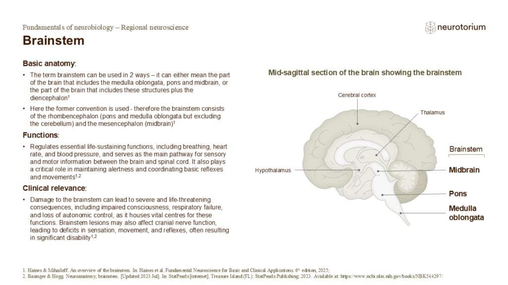
Brainstem
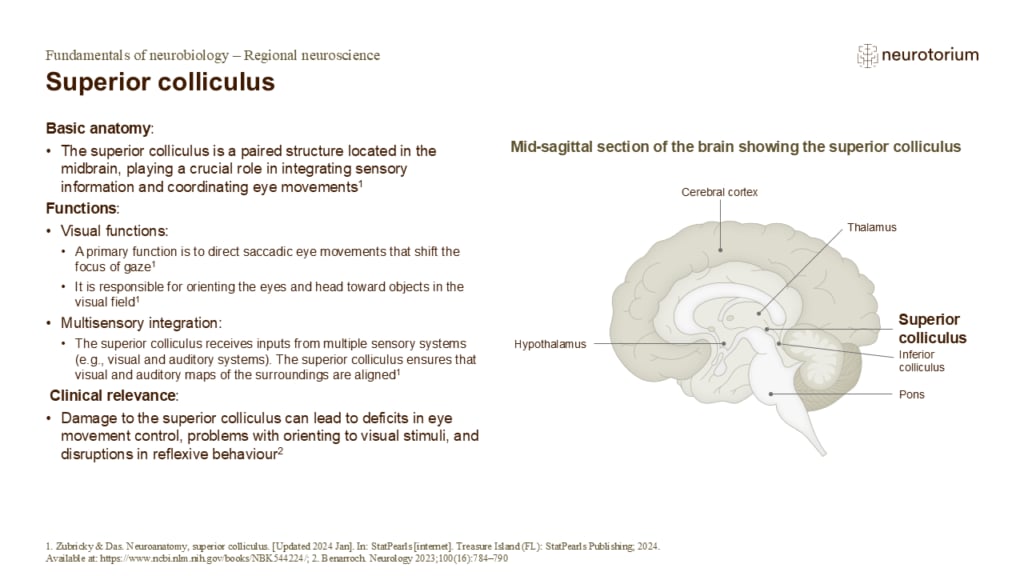
Superior colliculus
Structure continued:
- The superior colliculus is located on the roof of the midbrain, above the inferior colliculus (which is involved in auditory processing)1,3
- It is a layered structure with the superficial layers mainly processing visual input from the retina and visua…
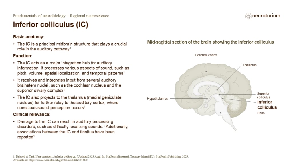
Inferior colliculus (IC)
The IC is divided into:1
- Central nucleus, which is the largest portion
- External cortex
- Dorsal cortex
The IC is involved in processing sound information before it is relayed to higher brain centres, particularly the auditory thalamus (medial geniculate body) and eventual…
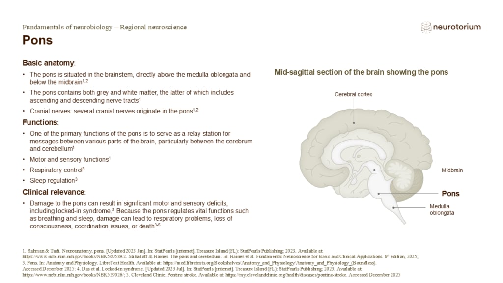
Pons
Several cranial nerves originate in the pons, including:2,6
- The trigeminal nerve (V) – responsible for sensation in the face and motor functions like biting and chewing
- The abducens nerve (VI) – controls the lateral rectus responsible for lateral eye movement
- The facial …
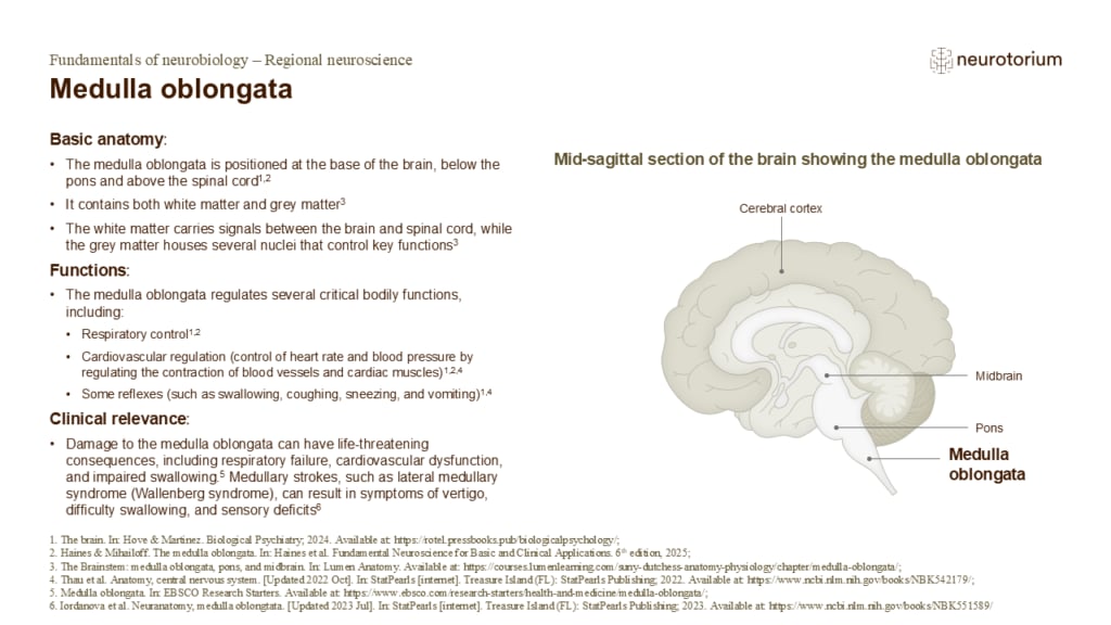
Medulla oblongata
The medulla oblongata is part of the brainstem, located just above the spinal cord. It plays a crucial role in controlling autonomic functions, such as breathing, heart rate, and blood pressure.1,2,4
Several cranial nerves emerge from or pass through the medulla, governin…
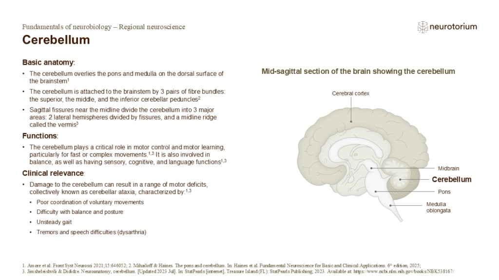
Cerebellum
The cerebellum, located at the back of the brain beneath the cerebral cortex, has a distinctive surface featuring many folds, or folia, that increase its surface area.1,2 It is divided into three main lobes (the anterior, posterior, and flocculonodular lobes) by transvers…
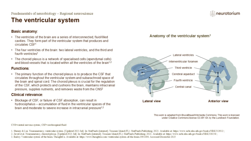
The ventricular system
The two lateral ventricles – one in each cerebral hemisphere are the largest of the ventricles. The third ventricle is a narrow midline cavity between the thalamus and hypothalamus. The fourth ventricle is positioned between the brainstem and cerebellum.1
References:
1. S…
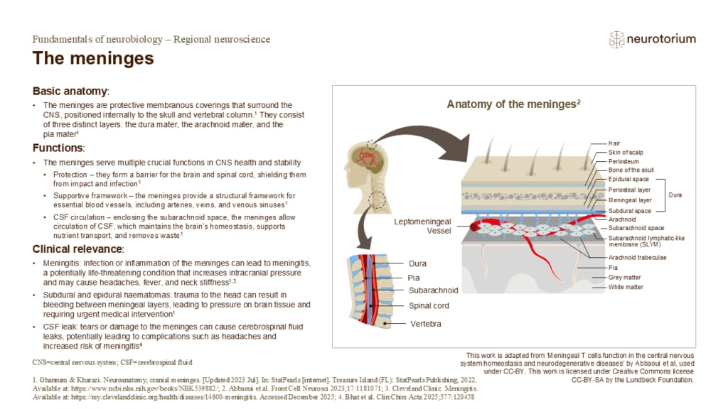
The meninges
The dura mater is a tough, thick layer composed of two sublayers, the periosteal dura (attached to the skull) and the meningeal dura (closer to the brain).1,2 The dura mater provides a protective layer for the brain and spinal cord.1,2
The arachnoid mater has a spider’s w…

The cerebrovascular system
The brain makes up approximately 2% of total body weight but consumes about 20% of the oxygen used.1 Oxygenated blood is supplied by the internal carotid and vertebral arteries.1 The internal carotid arteries enter the skull and branch into the anterior and middle cerebra…
Related content
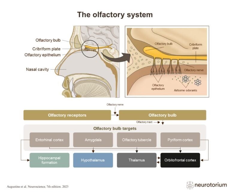
The olfactory system detects airborne odor molecules in the nasal cavity and transmits this information to the olfactory bulb. From there, signals are relayed to several brain regions involved in smell perception, memory, emotion, and behaviour.
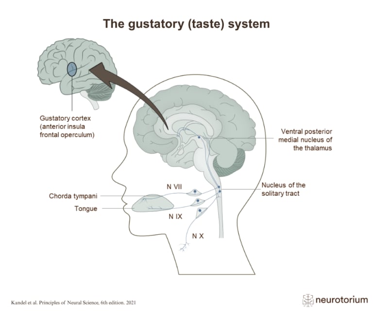
The gustatory system detects taste stimuli on the tongue and relays this information through brainstem and thalamic pathways to the gustatory cortex.
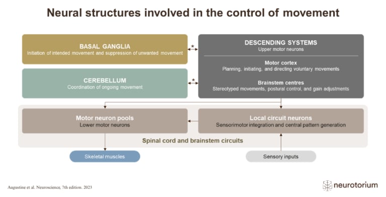
Movement is controlled by a network of brain and spinal cord structures that work together to plan, initiate, coordinate, and execute actions.